 Caridosuctor: Lund & Lund, 1984. C. populosum Lund & Lund, 1984.
| Sarcopterygii | ||
| The Vertebrates | Rhabdodermatidae |
| Vertebrates Home | Vertebrate | Vertebrate |
|
Abbreviated Dendrogram
Teleostomi
├─Neopterygii
└─Sarcopterygii
├─┬─Onychodontiformes
│ └─Actinistia
│ ├─Miguashaia
│ └─┬─Diplocercides
│ └─┬─Hadronector
│ └─┬─Caridosuctor
│ └─┬─Rhabdoderma
│ └─Coelacanthiformes
└─Rhipidistia
├─Dipnomorpha
└─┬─Rhizodontiformes
└─Osteolepiformes
├─Tristichopteridae
└─Elpistostegalia
├─Panderichthys
└─Tetrapoda
|
Contents
Overview |
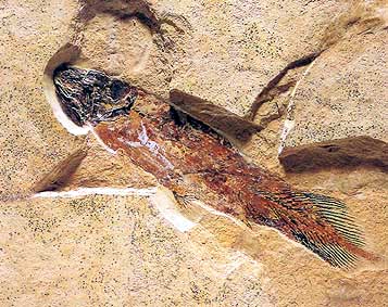
 Caridosuctor: Lund & Lund, 1984. C. populosum Lund & Lund, 1984.
Range: Mississippian (Serpukhovian) of North America (Montana) [1]
Phylogeny: Actinistia ::::::: Rhabdoderma + Coelacanthiformes) + *.
Characters: 8-22 cm [LL84]; premaxilla relatively large, with foramina for rostral organ [F98]; premaxillae may be separated by median rostral [F98]; rostrum and skull roof sparsely ornamented [LL84]; rostral region high & rounded [LL84]; skull table shields about equal length [F98]; two pairs. parietals above orbit, with posterior parietal much larger [F98]; tectal series with at least 12 elements [F98]; dermal intracranial joint simple [F98]; postparietal shield narrow, with straight posterior margin [F98]; supratemporal posterior margin even with posterior margin of postparietal [LL84]; intertemporal absent [F98]; 5 scale-like extrascapulars with straight posterior margin [F98]; antorbital large, with posterior rostral organ foramina [F98]; cheek bones broadly overlapping [LL84] [F98]; cheek completely covered with broad, tightly sutured bones [F98]; all cheek bones heavily ornamented [F98]; lacrimojugal not expanded anteriorly [F98]; large teeth on dentary, ectopterygoids and "precoronoid" (= anterior coronoids?) [LL84]; operculum articulates with supratemporal [LL84]; jaw long & shallow [F98]; jaw ornament minimal [F98]; Meckelian poorly ossified anteriorly [F98]; dentary without hook-shaped process, but slightly expanded posteriorly [F98]; dentary & splenial shallow [F98]; dentary pore present on splenial or between dentary & splenial [F98]; 3-4 coronoids [F98]; principle coronoid generally triangular, with curved anterior surface, straight posterior [F98]; angular with only small overlap area for gular [F98]; angular with small, rounded coronoid expansion [F98]; jaw articulation below posterior margin of skull [F98]; fused articular-retroarticular [F98]; premaxillae relatively large, with 3-4 robust teeth [F98]; large teeth on anterior coronoids [F98]; probably separate tooth plates on dentary [F98]; posterior coronoid with large teeth [F98]; body cylindrical [LL84]; body slender [F98]; 1st dorsal fin with ventral processes [LL84]; 2nd dorsal, basal plate elongated posteriorly with fin articulation on posterior end [LL84]; anal fin plate usually unossified, simple rod in body wall, located anterior to 1st hemal spine [LL84]; elliptical caudal fin [LL84]; caudal symmetrical with long middle lobe [F98]; cleithrum wide [F98]; extracleithrum large [F98]; pelvic plates very broad anteriorly, with 3 large & 1 small anterior processes [LL84] [F98] pelvic base with "strongly digitate medial process" [F98].
Links: Caridosuctor populosum the Bear Gulch web site); Quastenflosser - Lebende Fossilien small image of beautifully preserved specimen); New Page 12; 14. Caridosuctor ...; 群馬県立自然史博物館 - カリドスクトール・ポプロスム; Bear Gulch- An Exceptional Upper Carboniferous Plattenkalk article on Bear Gulch).
Image: Quastenflosser - Lebende Fossilien.
Notes: [1] The most common vertebrates of Bear Gulch. [LL84]
References: Forey 1998) [F98]; Lund & Lund (1984) [LL84]. ATW060119.
 Rhabdoderma: Reis, 1888. R. ("Coelacanthus") elegans Newberry 1856; R. ardrossense Moy-Thomas, 1937, R. ("Diplocercides") davisi Moy-Thomas, 1937, R. exiguum Eastman, 1902; R. huxleyi Traquair, 1881; R. madagascarensis Woodward, 1910; R. tingleyense Davis, 1884.
Range: Mississippian (Viséan) to Early Triassic of Europe, Ukraine and North America (central US) & Madagascar
Phylogeny: Actinistia :::::::: Coelacanthiformes + *.
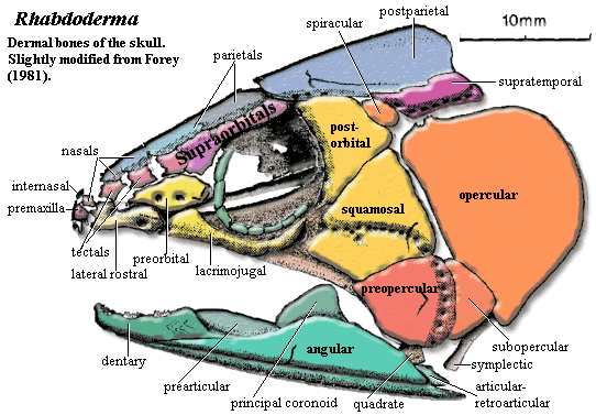
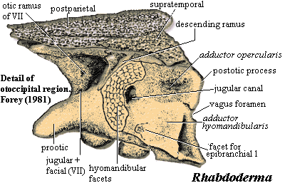
Characters: skull bones heavily ornamented (only near ossification centers in juveniles) except rostral bones [F98]; head with convex dorsal profile [F98]; Rostrum: rostral bones loosely associated [LL84]; premaxilla unornamented, bearing anterior foramen of rostral organ [LL84] [F98]; premaxilla large, notched for anterior nares [F98]; rostral bones largely in contact [F98]; Skull Table: median internasal present [F81] [F98]; parietonasal shield with 1 unpaired + 2 pr nasals & 2pr parietals increasing size posteriorly), about same length as postparietal [F81] [F98]; parietals contact with complex interdigitating suture [F98]; parietal descending process present, but weak [F98]; tectal series with at least 6 elements, including 4 supraorbitals, with tectals 4-5 larger than others [F98] postparietal lacks descending lamina [F81]; postparietals longer than broad [F98]; supratemporal with descending lamina along hyomandibular facets [F81]; supratemporal small, without postparietal lamina [F81]; postparietal shield with straight posterior margin [F98]; intertemporals absent [F98]; five extrascapulars [F81] [LL84]; 5 extrascapulars [F81]; lateral rostral small, with poorly developed ventral process [F98]; antorbital large, reaches orbit, & perforated by posterior openings of rostra1 organ [F81] [F98]; Cheek: cheek bones fit closely with overlap of preopercular on squamosal and squamosal on postorbital [LL84] [F81] [F98]; lacrimojugal tubular and not expanded anteriorly [F98]; postorbital triangular [F98]; posterior margin of postorbital is posterior of intracranial joint [LL84]; spiracular present, small, with scale-like ornament [F98]; squamosal largest cheek bone, triangular or pentagonal, with posteriorly-placed center of ossification [F98]; subopercular present [F81]; preopercular & subopercular arranged in same vertical plane [F98]; possible large median supraoccipital [F98] (dubitante);  Braincase: see additional figure; otoccipital portion of braincase completely ossified, without sutures, reaches skull roof (all primitive) [F81]; opisthotic with vertical crest bearing postotic process for epaxial trunk muscle attachment [F81]; opisthotic region with paired lateral depressions for (dorsal) adductor opercularis and (ventral) adductor hyomandibularis [1] [F81]; swelling over posterior semicircular canal continuous with a parampulary process bearing facet for epibranchial 1 [F98]; vagus (X) foramen and articulation for epibranchial 2 (lower right hand corner in image) just ventral to parampulary process [F98]; jugular canal, posterior entrance bilobed for passage of hyomandibular ramus of VII [F98]; sphenethmoid partially ossified [F81]; sphenoid condyles poorly developed & major element of intracranial joint is sliding articulation with processus connectens [F81]; antotic process well-developed [F81] antotic process narrow & only partly overlapped by parietal descending process [F98]; superficial ophthalmic presumably exits braincase under parietal descending process [F98]; profundus foramen just below antotic process [F81]; basipterygoid process absent or reduced to slight shoulder on processus connectens [F81]; processus connectens reaches parasphenoid ventrally [F98]; interorbital septum partially ossified [F98]; foramen for pituitary vein small, ventral to the optic foramen, at contact between basisphenoid & parasphenoid; [F98]; paired lateral ethmoids (ectethmoids) present, small & triangular [F81]; Palate: parasphenoid closely applied but not fused to braincase [F81]; palatoquadrate with three reinforcing spines (see image at ectopterygoid) [F81]; pterygoid covered with denticles [F81]; ectopterygoid and dermopalatines present [F81]; parasphenoid flat, laterally expanded anteriorly, and lacking dorsal process [F81]; quadrate not ossified [F81]; Mandible: mentomeckelian small [F98]; dentary small {F81]; dentary longer than splenial, posteriorly expanded but lacking hooked process [F98]; angular large, heavily ornamented [F81] [F98]; principal coronoid triangular [F81]; single proximal Meckelian ossification [F81]; Dentition: premaxilla bears 3-4 robust teeth [F98]; parasphenoid bearing teeth over entire surface almost to cranial fissure [F81] [F98]; dentary tooth plates present [F98]; coronoids with large teeth [F98]; prearticular covered with viliform teeth [F98]; five gill arches [F81]; branchial arches 1-3 with epibranchials, attached to single basibranchial [F81]; ceratobranchials with 3 rows of tooth plates, including gill rakers [F81]; ribs not ossified [F81]; all lepidotrichia unornamented [F81]; first dorsal fin with kidney-shaped endochondral support [F81]; caudal fin primary rays with one-to-one ratio to endochondral supports [F81] [LL84]; pectoral girdle ornament restricted to dorsal part of cleithrum [F81]; anocleithrum present [F81]; extracleithrum probably present [F81]; clavicles probably met on midline, or through hypothetical) interclavicle [F81]; triangular scapulocoracoid, largely unossified [F81]; pelvic fins inserting posterior to first dorsal [F81]; pelvic girdles suture across midline, bearing anterolateral extensions [LL84]; scale ornament of ridges converging on midline [F81]; air bladder with calcified walls [F81].
Links: New Page 12 (good image of fossil from Mazon Creek, Pennsylvanian of USA), CARBONÍFERO Peixes bad image of much better fossil); 川崎悟司イラスト集・ラブドデルマ life reconstruction); Fossil Tours & Displays another good photo of a very small specimen with a few interesting details of skull); The Coelacanth - IIDB some discussion with sketch of skeletal features)
Notes: [1] See figure for additional details of otoccipital region.
References: Forey (1981) [F81]; Forey (1998) [F98]; Lund & Lund (1984) [LL84]. ATW060119.
checked ATW060320