| Glossary | ||
| The Vertebrates | Po-Ps |
| Vertebrates Home | Vertebrate | Vertebrate | Bones | Time |
For most phrases beginning with directional words, e.g. "posterior," "dorsal," "external," etc., or some generic anatomical terms, e.g., "vena," look under the next word in the phrase. However, note that this convention is not used with complete consistency in this Glossary.
Pod''emnaya Formation that's not a typographical error. Its a Lochkovian exposure in Severnaya Zemlya. Sometimes spelled "Pod'emnaya."
Polar cartilage Two or more small cartilage bodies that form at the posterior end of the sphenoid region during development of the braincase. See The Braincase and figure, infra, at prechordal bars.
Polarity (of a character) Determination of polarity is the determination of which character state is primitive for a group, i.e. the character state of the ancestor of a monophyletic group.
Pollex pes digit 1; the big toe; 15 & 16 (if you count on your digits); this little piggy went to market; etc...
Polyphalangy the condition of having many phalanges. For example, aquatic reptiles have repeatedly evolved flippers from limbs by elongating their limbs with additional finger and toe bones.
Polyphyletic (of a group) a group which does not contain its own last common ancestor.
Polyphyodont having more than two sets of teeth in a lifetime, as opposed to diphyodont.
Polyprotodont having more than two lower incisors.
Polyspondyly [1] the condition in which the vertebrae have more than two centra. This condition is found in some dipnoans and holocephalians. It is also one of the few English words having 3 'y's, although it would not be difficult to construct others. For example: polycryoichthyodactyly -- the very common parental condition of having many frozen fish-sticks. [2] Also used in the special case of certain chondrychthyans which have calcified "rings" around the notochord at intervals which do not seem to correspond in a regular way to the placement of body segments.
POMCProopiomelanocortin.
Pondaung Formation Middle and Late Eocene of Myanmar. Terrestrial. Primates.
Popliteal area (or space or fossa) a depression on the ventral side of the femur, between the condyles, presumably related in some fashion to the popliteal nerve and/or artery.
Pore-canal system see cosmine.
Posongchong Formation Early Devonian (Pragian) of China (Yunnan). Onychodonts & galeaspids. Zhu & Janvier (1994).
Post (abbr.) posterior.
Post-dentary bones same as postdentary bones.
Postaxial radial see figure at Dipnomorpha
Postbranchial laminaof the cleithrum. See section on the elpistostegalian pectoral girdle.
Postcingulum a transverse cingulum running across the distal (posterior) face of an upper molar. If a lower molar, then postcingulid.
Postcristid in mammalian dentition, the ridge running around the distal end of a lower molar (the talonid side) from entoconid to hypoconid (i.e. with the hypoconulid more or less in the middle).
Postdentary bones collectively, the angular, surangular and articular bones. This term is used almost exclusively to refer to these bones in mammaliforms, in which they are not posterior to the dentary. It seems never to be used with reference to reptiles, in which the postdentaries are posterior to the dentary.
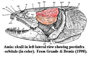
Postinfraorbital in fishes, dermal bones of the circumorbital series which are located posterior to the orbit and often elongated anteroposteriorly.
Postorbital articulation of the palatoquadrate. One of the possible dorsal articulations of the palatoquadrate in which the palatoquadrate articulates with a process on the posterodorsal part of the ethmoid.. See image at paratemporal articulation.
Postorbital bar A bony bar separating the orbit and temporal fenestra "temple").
Postorbital pila a dorsal or anterodorsal process of the basisphenoid from near the base of the basipterygoid process into the orbit. This process helps support the eyestalk, if present, and anchor the extrinsic eye muscles of the rectus series. Also called the basisphenoid pillar. See figure at rectus muscles; see also discussion and figures of the gnathostome orbit.
Postorbital Process A projection from either the jugal bone on the zygomatic arch, or the frontal bone, partially separating the orbit and the temporal fossa.
Posttemporal a paired bone of the sarcopterygian skull and shoulder girdle. The posttemporal attached to the skull anteriorly and also bridged the lateral extrascapular (dorsomedially) to the supracleithrum (ventrolaterally). See Eusthenopteron for an image of the whole series. The posttemporals are lost in all tetrapods.

Posttemporal fenestra in stem and basal tetrapods, the paroccipital process does not completely seal off the braincase. A space is left, generally bordered ventrally by the paroccipital process and dorsally by the tabular. The squamosal, the exoccipitals (if distinct from the paroccipital process), and the supraoccipital (if present) may also be involved. The fenestra defined by these elements leads to a shelf on the dorsal surface of the otic capsule.
Posttemporal foramen in some forms, notably basal archosaurs, the posttemporal fenestra is reduced to a small foramen. Normally, the foramen is bordered by the paroccipital process ventrally, and by some combinationof the parietal, squamosal and perhaps supraoccipital dorsally and laterally.
Postzygodiapophyseal lamina reinforcing ridge bone ridge in the vertebrae (normally, of sauropods) connecting the postzygapophysis with the diapophysis on the same side. See image at centrodiapophyseal lamina.
Praespiraculum in jawless fishes, an anterior head opening supposedly representing the gill slit of the mandibular arch, assuming that the mandibular arch was a functional gill arch at some point. The openings just medial or just lateral to the orbits in some very basal vertebrates are identified by some as praespiracula. Today, the more conventional interpretation is probably that these are nasal or nasohypophyseal openings.
Pragian Age Early Devonian. The second and middle age of the Lower Devonian, 412-400 Mya.
Preacetabular tubercle an anteriorly or laterally directed short process of the pubis located, as the name suggests, on the body of the pubis, anterior to the acetabulum. Known from, inter alia, various birds and lizards.
Preaxial radialsee figure at Dipnomorpha

Prechordal bars same as trabeculae. See also figure at right.
Precingulum a transverse cingulum running across the mesial (anterior) face of an upper molar. If a lower molar, then precingulid.
Precurrent preceding or, more specifically, the members of series e.g. vertebrae) which are more anterior to the subject member. "The prezygapophyses articulate with corresponding structures on the precurrent vertebra."
Prehensile Structures adapted for grasping or seizing by curling or wrapping around such as the tail of some American monkeys and opossums.
Prehension the act of seizing or grasping. For an interesting essay on tongue prehension vs. jaw prehension of food in lizards, see Foraging and Trophic Ecology.
Premaxilla a dermal bone of the facial series, including the anterior portions of the upper jaw and rostrum. See Bones: The Premaxilla for the gory details.
Premolar molariform replacement teeth mesial anterior) to the molars. "The fundamental distinction between premolars and molars is developmental: premolars developed from a secondary dental lamina and replace deciduous precursors (which may or may not erupt), whereas molars originate from the primary dental lamina and, by definition, neither replace precursors nor are replaced by successor teeth." Kielan-Jaworowska et al. (2002: 481).
Prepatagium a roughly triangular flap of skin on the wing of a bird or pterosaur which fills the space bounded by the humerus and ulna.
Presacral of vertebrae, the vertebrae other than the sacral (pelvic) and caudal (tail) vertebrae, i.e. the cervical neck) vertebrae and the dorsal (back) vertebrae, the latter including the thoracic (upper spine) and lumbar (lower back) vertebrae, if these regions are specialized (as in mammals).
Prespinal lamina reinforcing medial bone ridge in the vertebrae (normally, of sauropods) running down the anterior face of the neural spine. See image at centrodiapophyseal lamina.
Prespiracle see praespiraculum.
Pressure force per unit area.
Preural caudal in fish anatomy, the more anterior of the two "tail" regions, in which the hemal arches are joined distally to encircle the caudal artery & vein. More posteriorly, the caudal artery splits in two and runs along both sides of the spine. Preural vertebrae also lack hypurals. The two anatomical definitions (distally joined hemal arches & absence of hypurals) are used cumulatively or interchangeably.
Prezygodiapophyseal lamina reinforcing ridge bone ridge in the vertebrae (normally, of sauropods) connecting a prezygapophysis with the diapophysis on the same side. See image at centrodiapophyseal lamina.
Prezygoparapophyseal lamina reinforcing ridge bone ridge in the vertebrae (normally, of sauropods) connecting a prezygapophysis with the parapophysis on the same side. See image at centrodiapophyseal lamina.
Pridoli the Silurian is split into 4 epochs, rather than the usual three (i.e., Early, Middle & Late). The Pridoli is the latest Silurian. The Pridoli is not split into ages, so that it is often spoken of as the Pridolian Age as well as Epoch. It is sometimes (and probably correctly) spelled "Přidoli." The "Late Silurian" usually refers to the Ludlow plus the Pridoli.
Primary calcification a process apparently unique to neoselachian sharks in which calcification of the notochord occurs by secondary invasion of chondrocytes into the notochord itself. These cells deposit cartilage between the external and internal elastic membranes of the notochord. The cartilage is then secondarily mineralized. Prismatic? endochondral? perichondral? ossification -- dunno... And with all this secondary activity, why is it called "primary calcification"?
Primary feathers the flight feathers on the wrist, hand and fingers (carpometacarpus and phalanges) of a bird.
Processus alaris generic term for a wing- or fan-like process.
Processus connectens in Sarcopterygii, the posteriorly directed processes of the sphenethmoid section of the braincase which articulate with the otoccipital section. See image at Diplocercides.
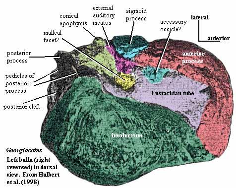
Processus tubarius in whales, by analogy with a structure on the human sphenoid, the anterior process of the bulla which supports -- actually forms -- the Eustachian tube. Generally, = anterior process of the bulla. Luo 1998). See also, image.
Procoelous a pattern of vertebral articulation in which the individual vertebrae are concave anteriorly and convex posteriorly. Opposite of opisthocoelous. lab7 photos
Procumbent slanted forward.
Procurrent inclined forward.
Procurrent ray one of a series of small, unsegmented rays on the dorsal and ventral edges of the caudal fin. Dictionary of Ichthyology.
Profundus nerve This nerve is known by a ridiculous number of names, most of which are variations on: V1 (i.e. branch #1 of the trigeminal or Vth cranial nreve), n. ophthalmicus profundus, or deep ophthalmic nerve. Note that the profundus is sometimes simply called the "ophthalmic nerve" because the term "superficial ophthalmic nerve" often refers to a branch of the facial (VII) nerve. However, the two often run together, making the term "ophthalmic nerve" ambiguous. Use of the English "deep ophthalmic" is also problematic. In some cases, the profundus splits into internal and superficial rami well forward of its exit from the braincase, in which case, the internal ramus of the nerve may be called the "deep ophthalmic," although both rami are part of the profundus. Finally, recent evidence suggests that the profundus is the same as the ciliary nerve in mammals.
The profundus exits the braincase with, or close to, cranial nerve V, the trigeminal nerve. In many sarcopterygians and actinopterygian fishes, the profundus exits together with the trigeminal and also enters into the semilunate ganglion with the trigeminal immediately outside the braincase. That is why the profundus was historically regarded as a branch of the trigeminal. That may or may not actually be the case. A good deal of theoretical wrangling depends on the answer, assuming that the question is meaningful (and it probably is). Some segmentalists associate the profundus with sensory inervation of the hypothetical pre-mandibular arch. Thus, they tend to argue that the profundus is a separate structure which is related to this hypothetical arch in the same way that the trigeminal (V) is related to the mandibular arch or the facial VII) to the hyoid arch.
Whether or not a premandibular arch ever existed, the profundus has certainly always been associated with sensory inervation of the most anterior parts of the head, and with the area around (but not in) the nasal capsule. This applies even in hagfishes, osteostracans, and other jawless forms.
In many osteostracans and gnathostomes, the profundus exits with, or just anterior to, the trigeminal and slightly posterior to the orbit. The profindus exit is located near the posterodorsal corner of the orbit in organisms with more or less laterally-placed orbits. The exit is frequently sheltered by a process of the ethmoid portion of the skull, such as a dorsal process for articulation with the upper jaw. From there, the profundus runs anteriorly across the interorbital septum, then dorsomedially over the orbitonasal wall, before plunging down into the area around the nasal capsules. In following this course, the profundus may enter and exit the bone/cartilage of the anterior braincase several times
Prognathous having the jaws projected anteriorly.Sometimes used to mean having only the lower jaw projected anteriorly.
Promaxillary fenestra a third anterior skull fenestra, in front of the antorbital and maxillary fenestrae.
Promontorium A bulge in the petrosal (otic capsule) of mammals marking the location of the cochlea in the inner ear.
Proopiomelanocorticotropin same as proopiomelanocortin.
Proopiomelanocortin ("POMC") an unusual hormone complex manufactured by the anterior lobe of the hypophysis. This complex is metabolized into four separate hormones: adrenocorticotropic hormone (ACTH), melanocyte stimulating hormone (MSH), encephalin and beta-endorphin.
Propalinal of jaw motions, using a back & forth front-to-back motion, as opposed to orthal, straight up and down.
Propatagium In bats and pterosaurs, thin web of skin that extends from the shoulder to the wrist anterior to the upper arm and forearm, and is fixed on the neck or skull.
Propodium the upper unit of the tetrapod limb, i.e., the humerus and/or femur. Same as stylopodium, in contrast to the lower unit, which is the zeugopodium or epipodium.
Propterygium fins are often attached to the torso by relatively large, columnar cartilages known as basals. Radials attach to the basals and extend towards the margin of the fin. Typically, there are three basals. In some cases, one of the basals becomes elongated, typically by adding segments bearing radials, and becomes the primary axis of the fin. When the anterior basal becomes extended in this fashion, it is referred to as the propterygium.
Protandrous sperm and eggs produced by same organ.
Prothecodont See Tooth Implantation.
Protocone the most lingual of the major cusps of the trigon on an upper tribosphenic molar; the mortar of the mortar & pestle formed by the protocone and talonid of a molar. See figure and Molars. See also figure at Mesostyle.
Protoconid the most buccal of the major cusps of the trigonid on a lower tribosphenic molar See figure and Molars. See also figure at Mesostyle.
Protoconule in mammalian upper molars. The nomenclature for small cusps in the mesiolingual (toward the tongue and anterior) region of upper molars is difficult. If the cusp is on the main body of the tooth, it is a protoconule. If it is a stylar cusp (derived from the cingulum) its a protostyle. If it takes the form of a ridge, it is an entoconule. The figure at entocone shows an upper right molar with both an entocone and a protoconule. Note that the protoconule, if present, will lie along the crista (ridge) connecting the protocone and the paracone, if such a crista is present. In any case it will be somewhere along a theoretical line between the two and probably mesiobuccal to the protocone. An entocone or protostyle is likely to be further out on the margin and mesiolingual to the protocone.
Protocristid in mammalian lower molars, the ridge joining the protoconid with the metaconid. This cristid probably has considerable physiological importance since this ridge forms a barrier which limits the motion of the opposing trigon to the talonid and thus prevents the opposing trigon "pestle" from slipping off the trigonid "mortar" -- the area where it is designed to occlude. This is particularly important to an animal which eats seeds, for example, which could easily cause the trigon cusps to slip out of occlusion before they could enter the talonid basin.

Protoloph in mammalian dentition, a cutting edge running generally along the mesial side of an upper lophodont molar. See image at lophodont and Molars.
Protostyle See protoconule or entocone.
Protraction in tetrapod locomotion, rotation of a limb anteriorly, in a horizontal plane. Opposite of retraction; and in contrast with adduction or abduction movement in a vertical plane) or rotation about the long axis of a limb. C.f. Humerus.
Proventriculus anterior stomach chamber.
Proximal closer to a reference point. If no reference point is indicated, then (usually) the center of gravity of an organism. Thus, the femur is proximal to the foot. "Proximal caudals" are the tail vertebrae close to the base of the tail. The "proximal tarsals" are the astragalus and calcaneum, because they are closer to the body than the distal tarsals (the navicular, cuboid, etc.). The opposite of proximal is usually distal.
Proximal articulation of the humerus, the articular surface of the proximal end of the humerus which articulates with the glenoid, and on which the humerus rotates.
Proximal carpals the upper wrist bones: radiale, intermedium, ulnare and (if present) pisiform. See figure at carpus.
Proximal tarsals the upper ankle bones, the astragalus and calcaneum. In mammals, these may be referred to as the tibiale and fibulare. The tarsal intermedium, if present, is also a proximal tarsal. See Figure at Tarsus.
Pseudohyal In batomorphs (rays and relatives), the ventral elements of the hyoid arch are reduced or absent. The hyoid rays coalesce into novel elements, termed pseudohyals, which functionally replace these elements. Compagno (1999a).
Pseudohypocone type of cusp that develops through cleavage of the protocone, unlike the true hypocone, which arises from the cingulum.
checked ATW030614