| Glossary | ||
| The Vertebrates | M |
| Timescale | |||||||
| Vertebrates Home | Vertebrate | Vertebrate | Bones | Essay Index | Geography |
For most phrases beginning with directional words, e.g. "posterior," "dorsal," "external," etc., or some generic anatomical terms, e.g., "vena," look under the next word in the phrase. However, note that this convention is not used with complete consistency in this Glossary.
M/m in mammaliform dentition, an upper (M) or lower (m) molar.
Maastrichtian the last age of the Cretaceous and of the Mesozoic Era, about 71.3-65.0 Mya.
Macrolecithal (of an egg) having a large amount of yolk. Macrolecithal development is the pattern of embryonic development characteristic of eggs with considerable yolk.
Macula (pl. maculae) L. macula = stain, spot, blemish. This root has no obvious relationship to the maculae, which are vaguely sac-like areas of the vestibular apparatus in the inner ear involved in the perception of linear acceleration, orientation in a gravity field, and low-frequency or high-volume sounds. See The Ear. Presumably the name comes from some histological property.
Maevarano Formation upK (Campanian?- Maastrichtian) of Madagascar.
Magnum in mammal osteology, one of the distal carpals, essentially a proximal extension of Mc III. Also referred to as the capitate. See image at unciform.
Malleolus rounded projections on either side of the distal end of the tibia which stabilize the tibia on the tibial trochlea of the astragalus. In other words, a couple of bone tabs at the end of the lower leg that help keep the leg properly oriented on the ankle joint.
Malleus the articular, when it is exapted as an auditory ossicle in mammals.
Mammalian jaw a jaw with a dentary-squamosal articulation, as opposed to the articular-quadrate articulation which was good enough for everyone else.
Mandagary Sandstone Late Devonian of New South Wales, Australia. The Mandagary Sandstone is the home of the Famennian Canowindra fauna, dominated by Bothriolepis, but with various other placoderms and sarcopterygians.
Mandible L. mandibula = jaw; from mandere = to chew. The lower jaw as a functional unit, regardless of which bones or cartilage make up the lower jaw in a particular organism.
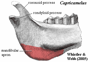
Mandibular apron a laterally thin expansion of the posteroventral section of the jaw in certain mammals.
Mandibular arch a hypothetical first gill arch which became the basis for the jaw. See Gill Arches.
Mandibular fenestra a fenestra or hole in the dentary -- a synapomorphy of crown group archosaurs.
Mandibular groove, internal same as Meckelian groove
Mandibular symphysis the area where the two halves of the lower jaw articulate.
Manse Burn Formation lwC of Scotland. Sequentially marine & nonmarine. Akmonistion, Deltoptychius, Denaea, Tristychius, actinopterygians, rhizodonts & osteolepids, coelacanth.
Manu- L. = hand
Manubrium [1] from the slight resemblance of the partially fused ribs to the fingers of a hand) anterior 3 segments of the sternum; more generally, the anterior portion of the sternum. See sternum. [2] the retroarticular process of the articular, when it becomes an auditory element of the middle ear in mammaliforms. A process of the malleus (articular) which attaches to the tympanic membrane and provides the insertion for the tendon of the tensor tympani.
Marble Most marble is made up of 90 percent or more of either calcite or dolomite; typically saccharoidal to relatively coarsely crystalline -- i.e., made up largely of grains a millimeter or more in greatest dimension.
Marl sediment composed of clay mixed with calcium carbonate calcite). Shell marl is marl in which the lime is in the form of invertebrate shells, i.e. biogenic (or calcareous) limestone.
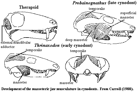
Masseter Gr. maseter = chewer. One of the few muscles named in the writings of Galen. The characteristic jaw muscle of therapsids which permits the longitudinal, grinding, motions of the jaw. The masseter is derived from the m. adductor mandibulae externus, which originates in the adductor chamber (in the temporal fenestra) and inserts on the coronoid process and internal surface of the lower jaw. See figure from Carroll (1988). In cynodonts, the adductor chamber is opened up as the dermal bones form a laterally bowed zygomatic arch. As a result, the origin of the external adductor mandibulae is split between the braincase and the medial surface of the zygomatic arch. The portion which remains on the braincase is referred to as the temporalis. The portion originating on the zygomatic arch now inserts in a fossa on the lateral surface of the dentary, the masseteric fossa, and is referred to as the m. masseter. In the later cynodonts, the masseter splits into deep and superficial elements. The superficial masseter is a new element which originates on the anterior portion of the zygomatic arch, below the orbit, and inserts on the posteroventral portion of the masseteric fossa. This combination of the temporalis with the deep and superficial masseters allows precise control of the jaws and, most significantly, control of mediolateral and anteroposterior movements, i.e. grinding. With this system in place, it became both possible and advantageous for cynodonts to evolve complex molar teeth designed for precise occlusion -- the hallmark of the mammalian condition.

Masseteric fossa The fossa for the masseter. In some therapsids, the coronoid process of the dentary bears a large external depression which accommodates the masseter -- an important muscle in closing and in grinding motions of the jaw.
Mastoid Gr. mastos = breast or nipple, and eidos = resemblance.
Maxilla the bone which normally forms the lateral upper jaw in osteichthyans, including tetrapods.
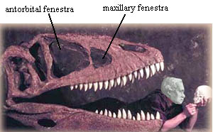
Maxillary fenestra a fenestra or hole which pierces the maxilla anterior to the antorbital fenestra. See figure. The maxillary fenestra is frequently inside the antorbital fossa, the large depression in the skull which includes the antorbital fenestra. For the terminally curious, no. There is no such dinosaur, and the head bearing integument is that of the noted German philosopher, Immanuel Kant.
Maxillary nerve the maxillary branch of the trigeminal nerve, or CN V2. The maxillary is a sensory branch which communicates sensations from the teeth, nasal sinuses, and in mammals) parts of the face lateral to the nose. The nerve is closely associated with the premaxilla.
Mazon Creek Pennsylvanian of Illinois, part of the Francis Creek Shale. Swamp, bayou & estuarine. Especially well known for chondrichthyan fossils.
Mc abbreviation for metacarpal.
Mecca Quarry Shale upC of the Mid-Western U.S. Part of the Cleveland Shale? The Mecca Fauna are a characteristic community of inshore marine or brackish water organisms which flourished during marine transgressions over swampland.
Meatus L. meatus = a channel or way. Thus the "external auditory meatus" is the channel for sound through the outer ear.
Mechanical Advantage the ratio of output force to input force.
Mechanoreceptor sensory cell or (less often) organ responsive to mechanical forces, such as water currents, touch, sound, or some moron with a really loud stereo in his car.
Meckelian bone the Meckelian cartilage is normally ossified (if at all) only at its ends, i.e. at the jaw symphysis and as the articular (in, e.g., reptiles). However, in sarcopterygians (other than crown tetrapods), and in a few other taxa, it may ossify completely -- in which case it would be rather lame to continue referring to it as the "Meckelian cartilage," as if we hadn't noticed the difference. When the Meckelian is ossified anteriorly, it is also referred to as the "mental bone."
Meckelian cartilage The cartilage formed in the mandibular process of branchial arch I; the primitive lower jaw, derived from the ceratal element of the hypothetical mandibular arch. See Gill Arches, Meckel's Cartilage, Gnathostomata.
Meckelian foramen in some sarcopterygians, (including many temnospondyls and lizards), the Meckelian cartilage is not completely surrounded by dermal bone, but lies in an open fossa visible on the medial face of the lower jaw.
Meckelian groove same as Meckelian fossa, foramen, or canal, but usually refers to tetrapods.
Medial referring to the midline of the body (or, occasionally, the midline of some other reference point). For example, the heart is medial to the ribs; and a biped must place its limbs medially when walking. Note, however, that a medial view is a view from the midline, looking outward. Opposite of lateral. The terms axial and radial are usually (not always) equivalent to medial and lateral, respectively. See medial for a discussion of related terms.
Medial centrale One of the distal carpals. See Figure at carpus.
Median in the middle. When used correctly, this is a more general term than medial, which refers to a specific axis. A classic example of confusion in these terms (possibly my confusion) is briefly discussed at Hadrosauroidea. The problem there is is the meaning of the phrase "medial carina" on a tooth. A median carina would be a keel up the middle of the tooth (on the inside face, the outside face, or both). A medial carina would be a keel on the inside, or lingual, face of the tooth. A mesial carina would be a keel on the anterior face of the tooth. So, it really does make a difference.
Median eustachian foramen in crurotarsans, a foramen on the ventral surface of the basioccipital and/or basisphenoid which communicates with the pharynx via the eustachian tubes. It is also referred to as the medial pharyngeal recess.
Medial pharyngeal recess same as median eustachian foramen. Alcober (2000), citing a paper by Witmer (1997) which we have not reviewed.
Median tuberosity a bulge on the proximal femur which is an attachment site for ligaments (ligamentum capitis femoris) binding the femoral head in the acetabulum. See figure at femur.
Medulla L. medulla = marrow. Used for the non-cortical (peripheral) part of some organs, such as the kidney and adrenal.
Melon organ in odontocete whales, a complex pocket of connective tissue and unusual, frequently toxic, fats located on the concave "forehead" formed by the maxilla between and anterior to the orbits. The melon organ is believed to focus ultrsound clicks used for echolocation. See discussion at Cetacea.
Mental [1] relating to the mind, from L. mens (pl. mentes) = mind; [2] relating to the chin, from L. mentum (pl. menti) = chin. I understand that many 3rd declension Latin nouns, such as mens, were taken over from the Romans' early mentors, the Etruscans. Presumably this is why there are two, very similar Latin words here with unrelated roots and meanings. This hypothetically Etruscan root is quite possibly related to Greek terms such as mania. Whoever the Etruscans were -- a mystery at least as deep as the origin of vertebrates -- they seem to have had Greek and Semitic influences. [3] relating to Meckel's cartilage. Thus the mental bone is an anterior ossification of Meckel's cartilage.
Mental foramen a pit, hole, or channel in the mandible (usually in the dentary) leading to the Meckelian cartilage or bone.
Mental groove in snakes, on the underside of the head, an anterior mental scale generally is followed by large, paired chin shields and smaller gular scales. Most snakes have a longitudinal mental groove between rows of chin scales.
Mentomeckellian the mental bone, a bone developed by ossification of Meckel's cartilage in the region where the two halves of the lower jaw meet (the mandibular symphysis).
Meristic of a character, having a number of parts, or divided into serially repeated, countable features (e.g. the rays in a fin, the myomeres of an eel larva, the rakers on a gill arch, etc.). From FishBase.
Mesenchyme See Early Development Terms.
Mesial [1] relating to dentition, closer to the axis of symmetry passing (vertically) through the front (symphyses) of the jaw, as opposed to "distal." In these Notes, "anterior" may be used instead of "mesial." Since we are usually discussing molars, the difference is immaterial. See medial for a discussion of confusingly similar terms. [2] In British, the word seems to be synonymous with medial, as in the opposite of lateral. This is unbelievably confusing when English-educated writers are discussing jaws and dental matters.
Mesocuneiform one of the distal tarsals. See ectocuneiform for image and explanation.
Mesodactyl same as mesaxonic.
Mesokinetic joint joint along articulation between frontals and parietals. SQUAMATES.
Mesolecithal (of an egg) having a moderate amount of yolk. Mesolecithal development is the pattern of embryonic development characteristic of eggs with moderate yolk.
Mesomere (a) a mesodermal component; see Early Development Terms. (b) the median series of bones supporting the pectoral fin of sarcopterygian fish. See Dipnomorpha. In the pectoral fin, the first mesomere is the humerus, the second the ulna, and the third probably the intermedium. The radius is the first preaxial radial. All other homologies are suspect.
Mesoplastron one of the dermal bones in the plastron of turtles.
Mesopodium the bones of the ankle and wrist, that is the tarsals and carpals (both proximal & distal), variously known as astragalus, calcaneum, fibulare, intermedium, tibiale, ulnare, intermedium, and radiale, and all centralia. See figure at metapodial
Mesopterygium fins are often attached to the torso by relatively large, columnar cartilages known as basals. Radials attach to the basals and extend towards the margin of the fin. Typically, there are three basals. In some cases, one of the basals becomes elongated, typically by adding segments bearing radials, and becomes a primary axis of the fin. When the middle basal becomes extended in this fashion, it is referred to as the mesopterygium.
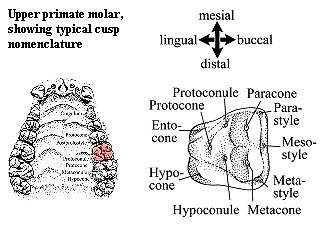
Mesostyle in mammalian dentition, one of the stylar cusps along the buccal margin of an upper molar. The mesostyle is, appropriately enough, the one in the middle. See figure.
Mesotarsal jointan ankle joint that passes between the proximal and distal tarsals, as in dinosaurs. Compare crurotarsal.
Mesothelial relating to the tissue lining body cavities.
Mesozoic Era the Triassic, Jurassic and Cretaceous Periods (248-65.0 Mya).
Metacarpus Gr. meta = after or beyond, and karpos = wrist. The wrist bones between the distal carpals and the finger bones (phalanges). See Figure at carpus.
Metacone the most posterior & buccal) cusp of the trigon on an upper tribosphenic molar. See Molars.
Metaconid the most linguodistal cusp of the trigonid on a lower (tribosphenic) molar. See Molarsor Teeth, diastema lengths.
Metaconule in mammalian dentition. Apparantly, this is the same as the hypoconule, that is, a small cusp on an upper molar lying near the line between the protocone (the lingual main cusp) and the metacone the distal main cusp). If the line is marked by a ridge, the ridge is the postprotocrista. By "main cusp," I mean one of the large cusps defining the vertices of the trigon. See figure at mesostyle.
Metaconulid the most distal cusp of the trigonid. See Molars.
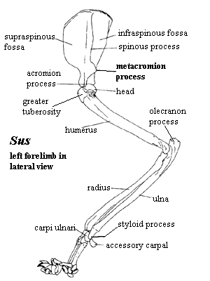
Metacromion process in mammals, a lateral, posterolateral or dorsolateral process of the coracoid portion of the scapulocoracoid, appearing as a outwardly-produced extension of the spinous process near its ventral (humeral) end. It frequently bears a further, angled projection at its highest point. In the cat, the metacromion serves as an attachment for the middle slips of the m. trapezius (i.e. the m. acromiotrapezius), as well as the insertion point of the m. levator scapulae ventralis, which originates on the atlas. A particularly emphatic (and very French) shrug might be summarize the motions involved.

Metakinetic joint "Joint occurs where parietal and supraoccipitals meet (metakinetic articulation)" PPT Slide. A joint "between the braincase and skull roof." Lee et al. (1999: 656). "In squamates other than snakes, the metakinetic axis passes through the tips of the paroccipital processes and their suspension from the supratemporal and, in some taxa but not Varanus, the squamosal." Rieppel & Zaher (2000: 492). At any rate, a joint at the intersection of the skull table and occipital region. An animal with a metakinetic joint is said to exhibit metakinesis.
Metaloph in mammalian dentition, a cutting edge running generally along the distal side of an upper lophodont molar. See image at lophodont and Molars.
Metalophid in mammalian dentition, a cutting edge running generally along the distal side of the trigonid on a lower lophodont molar. See image at lophodont and Molars.
Metamere a body segment
Metameric characterized by serial repetition of segments.
Metaphysis the sub-terminal, actively growing section of long bones. As opposed to the diaphysis and epiphysis q.v.).
Metapodials the metatarsals and/or metacarpals. Legs, Feet, and Locomotion at Animal Diversity Web:. The term sometimes includes the phalanges as well.
Metapophysis in some mammals, the articular surfaces of the vertebrae are on the neural arches, rather than on the centra. The metapophyses are the anterior articulations, analogous to the prezygapophyses. Frequently both structures are present and in close proximity. The corresponding posterior articulations are the anapophyses.
Metapterygium fins are often attached to the torso by relatively large, columnar cartilages known as basals. Radials attach to the basals and extend towards the margin of the fin. Typically, there are three basals. In some cases, one of the basals becomes elongated, typically by adding segments bearing radials, and becomes a primary axis of the fin. When the posterior basal becomes extended in this fashion, it is referred to as the metapterygium. Shubin (1995). Sometimes this results in an elongated, thin, extension of the fin, which is referred to as a metapterygial whip. The individual segments along the metapterygial axis are referred to as mesomeres, and the side-branches (on one side or both are referred to as radials. A distinction is often made beween radials (or other features) which occur on the leading and trailing sides of the axis, and these structures are referred to as being preaxial and postaxial, respectively.
Test yourself: it should now be possible for you to translate such classic ichthyo-babble as "the third mesomere bears two elongate preaxial radials."
Metapterygoid the portion of the palatoquadrate (primitive upper jaw) which actually articulates with the braincase to form the dorsal and/or basal articulation(s). Long explanation: the upper jaw in basal fishes consisted of a large, at least partially ossified, cartilage called the palatoquadrate. The upper jaw can be attached to the skull in a number of ways, but the most common attachments involve dorsal and/or ventral articulations (which may or may not be moveable joints), usually located just behind the orbit. This is accomplished by means of some postorbital process of the ethmoid region of the braincase dorsal) and/or a basipterygoid process (ventral). The metapterygoid is the part of the palatoquadrate which holds on to these processes. In tetrapods, this arrangement is largely replaced by dermal bone (the pterygoid) except for the dorsal articulation itself, which is formed by a small remnant of the metapterygoid called the epipterygoid.
Warning: in actinopterygians, metapterygoid has another meaning, for which we have been unable to pin down a reasonable definition.
Metastyle a stylar cusp near the buccodistal corner of an upper tribosphenic molar. See figure at mesostyle.
Metastylid a small, presumably stylar, cusp on the lingual edge of a lower tribosphenic molar, typically located just distal to the metaconid and more or less in the break between the trigonid and talonid. Appears to be the same as the tuberculum intermedium. See Teeth, diastema lengths.
Metatarsus Gr. meta = after or beyond, and tarsos = instep (actually, the ankle). The Metatarsals are the long bones inside the foot (in humans), between the ankle bones (distal tarsals) and the toes (phalanges).
Metataxon In cladistics, a group of species or specimens which is assumed in advance to constitute a clade for purposes of establishing relationships among other groups (not sure about this one). This is frequently necessary in two situations. (a) If the existing material is scrappy, and it may make better sense to combine odds and ends of material which is very likely related as one metataxon, rather than use numerous taxa with missing data. (b) If the taxon contains one or more derived groups which exhibit reversals of character states which were originally synapomorphic.
Metautostylic a type of jaw suspension characteristic of tetrapods other than mammals) in which the lower jaw is supported by the quadrate; that is, the basic quadrate-articular jaw articulation.
Methyostyly "Hyostilic jaw suspension is found in elasmobranchs. The jaw is formed by the palatoquadrate and Meckel's cartilage, and they are suspended from the hyomandibular element that attaches to the cranium. Methyostylic jaw suspension is a derivation of hyostilic suspension in that the hyomandibular still plays a role in jaw suspension, but in this case the original elements have been replaced by dermal bone, and the hyomandibular is a reduced element at the jaw joint. Methyostilic suspension is found in Osteichthyes." Midterm_key_2001.pdf
Metotic Fissure an embryonic fissure between the otic capsule and the arches which develop into the occipital bones. The fissure may persist into adulthood. It is also referred to as the "lateral otic fissure." The metotic fissure opens into the vestibular fontanelle, if the fontanelle is present. Johanson et al. (2003). Otherwise it opens to the metotic foramen. See the inner ear of reptiles.

Metotic foramen a foramen by which the metotic fissure may open to the outside. See the inner ear of reptiles. Strictly speaking, the metotic foramen refers to an undivided opening which serves both as the lateral opening of the recessus scala tympani (i.e. it is the fenestra pseudorotunda) and as the exit for various cranial nerves (i.e. the vagus foramen). More broadly, it refers to the region in which both (if separate) are found.
Microlecithal (of an egg) having little or no yolk. Microlecithal development is the pattern of embryonic development characteristic of eggs with little or no yolk.
Microsquamose having very small scales.
Microvillus (pl. microvilli). See villus.
Milk River Formation middle Cretaceous to Late Cretaceous (Campanian?) of Alberta, Canada. Multituberculates. Esociformes.
Mixopterygium the pelvic copulatory "clasper" of male chondrichthyans. Sometimes spelled mixipterygium.
Modesto Formation Late Carboniferous of Illinois
Molar L. mola = a millstone. Hence, a grinding tooth.
Moment Arm In a rigid system, the distance between a reference point and the point at which a force is exerted on the system. See torque.
Monoecious having one sexual form. Not asexual, but every individual produces both gametes.
Monophyletic descended from a single common ancestor within the group being studied. The term is also used in a slightly different sense. A "monophyletic group" usually refers to a clade, i.e. an organism and all of its descendants. Thus Sinraptor and Allosaurus are (very likely) monophyletic within Allosauroidea. However they are not, by themselves, a clade, because they do not include all descendants of their last common ancestor.
Monospondyly the condition of a spine having one vertebral centrum per segment.

Monte San Giorgio Middle Triassic locality in northern Italy and southern Switzerland, south of Lake Lugano. The locality is particularly well-known for pachypleurosaurs. Expeditions from the University of Zurich began excavating exposures here in the 1930's. Since this was Switzerland, and since no one ever told them to stop excavating, they continued to dig with ceaseless diligence for 40 years. Sander (1989). Mercifully, someone seems to have realized what was going on and put a stop to the madness before the entire southern Alpine Massif was carved into tidy slabs, carefully labeled, and deposited in the cavernous drawers of the Zurich University Museum. Note that the valley now filled by Lake Lugano follows the line of the Monte San Giorgio locality exactly and lies just a little to the north. This raises a truly disturbing question: is Lake Lugano really a natural phenomenon -- or might it represent the scars of other extended fieldwork by these implacable Swiss paleontologists?
Montehermosan Age South American Land Mammal Age corresponding to the Early Pliocene.
Moran Formation Wichita Group, Asselian or Early Sakmarian (Cisuralian: Permian: Paleozoic) of Texas. Lupeosaurus, an edaphosaur (Eupelycosauria: Synapsida) (Romer & Price, 1940) and Romeria, a captorhinid (Benton & Donoghue, 2007). Marginal marine sediments of thin limestone beds, massive shales, and lenticular channel fill sandstone. Includes shellbanks of the mollusk Myalina.
Morphotype a theoretical construct of the primitive form of a taxon. A hypothetical generalized form, having all the synapomorphies of a group and no specializations.
Morrison Formation an enormous upJ (Tithonian?) formation with various exposures. The formation stretches from northern New Mexico up to southern Canada and takes in the entire states of Colorado and Wyoming (although it is naturally overlaid in most places). The Morrison has two principal members, the Salt Wash (lower) and Brushy Basin upper).
Mosaic (a) having a mixture of features characteristic of more than one lineage -- which usually means that some of the features are plesiomorphic after all (b) made up of small pieces, as a mosaic tile.
Mosch- Greek root for calf.
Mt abbreviation for metatarsal.
Mustersan South American Land Mammal Age roughly corresponding to the Middle or Late Miocene.
Mylohyoid groove obsolete term for Meckelian groove
Myodome a dome-like space in bone or cartilage which is presumed to be occupied by muscle in life.
Myomere A unit of segmented muscle separated from contiguous units by a connective tissue, particularly the embryonic muscles blocks formed by the mesodermal myotome derived from each somite in early development. Thus the myomeres mark the embryonic segmental pattern originally established by the somites. See, generally, Early Development Terms.
Myotome a myomere enervated by one spinal nerve; a voluntary muscle segment. In embryology, see Early Development Terms.