 Actually, this is a kind of Cubist perspective, (see Figure 2, reproduced by permission), since we are attempting to view the occlusal surfaces of both upper and lower teeth at once.
| Bones: Teeth | ||
| The Vertebrates | Molars |
| s | ||||||
| Vertebrates Home | Vertebrate | Vertebrate | Bones | Time |
|
Bones
|
Teeth
|
While it will be necessary to go into this subject in more detail at another time, this section will serve for now as a sort of temporary filling: a place to put some molar nomenclature for the tribosphenic molar until a fuller discussion is possible. For purposes of the following discussion, almost all other puns have been omitted and are left as an exercise for the reader.
Some conventions: for purposes of these notes, the orange/tan color is used for upper molars and the blue, for lower molars. Anything dealing with the lowers has the suffix -id. This gets carried to extremes in some literature, and I will try to avoid it. Cusps are the elevated points of teeth, and are shown as pink circles. The addition of -ule to a cusp name means a subsidiary cusp. Thus, the posterior-most cusp of the trigonid is the metacone, and a subsidiary cusp of the metaconid would be the metaconulid. Cristae are the ridges joining the cusps, and are shown (when present) as pink lines. Shearing surfaces are shown with a pattern of short grey lines, while broad crushing surfaces are stippled in grey. All figures are shown in a sort of occlusal view, meaning we are looking down on the business end of the tooth, similar to the last thing a prey animal ever sees. Actually, this is a kind of Cubist perspective, (see Figure 2, reproduced by permission), since we are attempting to view the occlusal surfaces of both upper and lower teeth at once.
The basic reversed triangles pattern for molars is shown schematically in Figure 1. This is the sort of dentition seen in Kuehneotherium, as well as symmetrodonts and other basic Mammalian types. Unfortunately, molar nomenclature was developed for therian mammals, and some of the terminology turns out not to work very well at this fundamental level. The basic triangular units with three cusps are the trigon and trigonid. The metacone is the posterior cusp. The A and B cones were originally thought to be homologous with the protocone and paracone, respectively, of therians (see below). This turns out not to be the case: hence the use of letter designations.
As the trigon and trigonid meet, the slide past one another, shearing the food item on the edges of the molars as shown. Depending on the shape and length of the cusps and the sharpness and position of the cristae, the molars may also pierce, at the tips of the cusps, or even slice like the teeth of ornithischian dinosaurs. However, the principle effect is shear, since neither the cusps nor the cristae actually occlude with anything. Instead, they are designed to slide past their opposite numbers.
 The "reversed triangles" molar, for all its elegance, was a somewhat limited system. Some foods resist shearing: seeds, bones, and nuts, not to mention chewing gum. Evolution, fortunately, developed a method for overcoming a potential culinary limitation to tofu, blanc mange, and strained peas by elaborating the upper molars lingually and creating a low posterior extension of the lower molar, the talonid. The margin of the talonid is surrounded by a series of cusps ("cuspids," actually, since this is a lower jaw molar) which enclose a talonid basin. The protocone and, if present, the hypocone of the upper molar occlude directly with this surface. Even when not reducing peanuts to peanut butter, this mechanism serves to guide the rest of the jaw into a more precise occlusion. The enlarged surface and finer occlusion also permitted the development of complex series of subsidiary cusps and other accoutrements. These include not only subsidiary cusps on the tooth surface, but ancillary stylar cusps on the cingula, the mineralized ridges at the base of the lingual face of the molar.
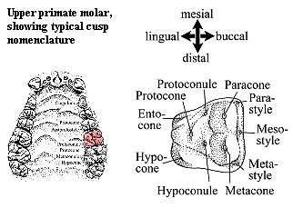
Evolution never comes without a price; and, in this case, the price included an explosion of dental nomenclature which can be deeply unsettling -- even to hardened paleontologists who can normally chew up a Greek root faster than you can say fluorophenylphthaline. Thus, we will dodge the fine points (and, to be sure, the fine pointids, pointules, etc.). The pages of Introduction to Teeth at Animal Diversity Web are strongly recommended for more, and certainly better illustrated, information and some very good real-life examples. However, there are a few important variations on the basic plan which require explanation.
The bunodont dentition of, for example, pigs (see Ungulate teeth, More on Artiodactyla) follows the basic pattern and consists of low, rounded cusps. It is characteristic of fairly unspecialized omnivores.
Ruminants need only grinding and cutting teeth. Thus they tend to revert to the trigonid form, but without cusps, so that the work is done by curved ridges: lophodont dentition. A loph is a ridge of enamel. The intervening dentine wears faster than the enamel resulting in distinct, fairly sharp contact surfaces. In selenodont molars, the enamel ridges form characteristic crescent shapes. See More on Morphology of the Artiodactyla (figure at bottom of page); selenodont.jpg. In extreme cases the molar dentition consists of close packed lophodont teeth with a single long lingual-buccal ridge: loxodont teeth. See recently2 mammoth teeth).
 Zalambdodont molars have an upper molar characterized by a V-shaped crest along the margin (ectoloph). At the apex of the V (on the lingual side of the tooth) is usually the paracone (sometimes fused with the metacone). This looks odd, since the paracone is typically at the buccomesial corner of the molar. However, animals with zalambdodont molars have offset jaws, so that the ectoloph is occluding with the hypoflexid on the lower molar, just as the paracone typically does. Asher et al. (2002). The crests of the ectoloph run to stylar cusps on the labial side of the tooth. The protocone is typically absent. See The Diversity of Cheek Teeth at Animal Diversity Web. A dilambdodont upper molar is similar except that the ectoloph is W-shaped. The metacone and paracone are at the (lingual) base of the 'W.' Crests run from these cones to buccal stylar cusps and form the arms of the 'W.' In addition, the molar has a low shelf, lingual to the rest of the tooth, with a small protocone. See, The Diversity of Cheek Teeth at Animal Diversity Web.
In some carnivores, one or two of the cheek teeth may be carnassials, specialized blade-like cutting teeth. La Brea Tar Pits.
Since the time the earlier part of this Note was written, our tastes in dentition have become a bit more refined, requiring a little more coverage and something a bit more substantial to chew on. To the right is an upper left molar adapted from de Muizon & Cifelli (2000) which will do for the purpose. I have deliberately not made the diagram more colorful or easier to follow than the original precisely so that we can learn to follow the usually obscure and unhelpful tooth diagrams in the literature.
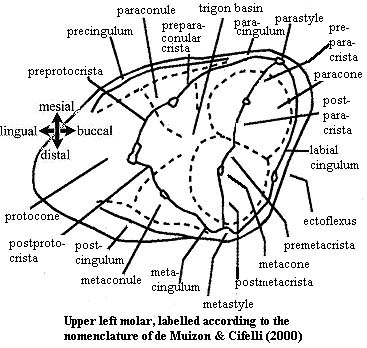 First, recognize that this is a left molar. We are looking at in occlusal view, which in this case means the same as a palatal perspective. This reverses the usual directions. That is, the tooth would be on the right of the diagram if the entire mouth were shown. Accordingly, lingual (towards the tongue) is on our left and buccal on the right.
Cones & conules: The easiest place to start is usually with the two major buccal cusps, the paracone (mesial or anterior) and metacone distal or posterior). Each of these often has a substantial conule lingual to it, the paraconule and metaconule, respectively. Lingual to all these other cones is the protocone. The trigonid basin lies in the center of all of these cones and conules. (This particular molar lacks a hypocone. If one were present, it would lie on the ridge created by the postcingulum.)
Cristae: The major buccal cusps also define a line which is usually marked by a crista. The regions of this crista are named for the cusp nearest them. Thus, the postparacrista is the region behind (distal) to the paracone. Distal to this segment is the premetacrista, and so on. The same convention is used for the cristae connected to the protocone. The system breaks down somewhat for the cristae associated with the conules, but in theory it remains the same.
Stylar cusps and cingula: A labial (i.e. buccal) cingulum runs around the buccal side of the upper molar, and stylar cusps are often associated with the points at which the preparacrista and postmetacrista intersect the cingulum. Not surprisingly, these are the parastyle and metastyle, respectively. Frequently, an additional stylar cusp (not shown) occurs between them. This is the mesostyle. Branches of the cingulum curve around outside the trigon on its mesial and distal sides. This are called, with impeccable logic, the precingulum and postcingulum.
That is essentially all we normally have to deal with on the upper molar. A right lower molar is shown in the next figure. We are now looking down on the tooth i.e. not a palatal view) but the subject is a right tooth, rather than left. These two effects cancel, so the chirality is the same as in the last image.
 Conids and conulids: Unfortunately, the nomencalture is not quite as logical for lower teeth, which are a bit more complex. By analogy to the upper molar, we can orient ourselves with the paraconid and metaconid. Since this is a lower molar, these cuspids are located on the lingual, rather than the buccal side. That's why the tribosphenic molar is referred to as "reversed triangles." Since there's a large talonid (particularly large in the example), the metaconid and paraconid tend to be found in the linguomesial quadrant. Somewhere buccal to these conules will lie the protoconid, and the three will define the trigonid, with the trigonid basin in the center. Truthfully, even the trigonid cusps can be very hard to identify. For example, look at Figure 12.2m on one of the best mammal tooth sites. This molar has no paraconid and has a very large entoconid. Consequntly, the entoconid and metaconid are likely to be misidentified as the metaconid and paraconid, respectively. The take-home lesson is that one should double check for the constricted "waist" between the trigonid and talonid. (Not infrequently, the lower third molar (m3) will appear to have two waists and three compartments. This is actually very helpful because, in that case, the cusp in the most distal section can safely be identified as the hypoconulid.)
Assuming we can correctly identify the trigonid, the next step is to identify the hypoconid in the buccodistal quadrant. As previously noted, there is a chance of confusion in nomenclature since the names of the hypoconid and entoconid are sometimes reversed. Nevertheless, the large cusp in the buccodistal quadrant is usually easy to find. The hypoconulid can then be distinguished from the entoconid (if both are present) because the hypoconulid is near the distal margin, or even set off distally from the main body of the talonid, while the entoconid is near the lingual margin.
Cristids: On the right lower molar, the cristids of the trigonid are named as if they were a clock face. That is, the cristid clockwise from the paraconid is the paracristid. The cristid clockwise from the protoconid is the protocristid. This is, of course, reversed on the left lower molars. Perhaps more frequently, the entire cristid around the trigonid is referred to as the precristid. On the talonid, the crista obliqua is often of considerable interest. Its position is easily determined from its origin at the hypoconid. Again, the entire cristid associated with the talonid is often referred to as the postcristid.
Cingulid and stylids: Unfortunately, these are so variable that I have been unable to get a good handle on them at this point. Accordingly, discussion is deferred for the moment.
 5. Lophodont Dentition
Ruminants and certain similar forms present special problems of nomenclature. Here, the cristae joining the cusps frequently become the primary dental surfaces. The cusps themselves may no longer be evident as separate structures. In many cases, the overall form of the cutting surface becomes so strongly simplified that no special nomenclature is necessary. However, intermediate states exist in which the form and placement of the lophs assume phylogenetic importance. The nomenclature used for lophodont forms is not always consistent. Purely by way of example, the image at left shows the nomenclature used for notoungulates, an extinct group of ungulates endemic to the early Cenozoic of South America. ATW021228.
Useful Web Sites
DENTITION DES VERTEBRES
Teeth, diastema lengths (Best on the Web)
Animal Diversity Web: Introduction to Teeth
checked ATW030403
