 Archenteron: same as gastrocoel.
| Glossary | ||
| The Vertebrates | Early Development Terms |
| Vertebrates Home | Vertebrate | Vertebrate | Bones | Time |
 Archenteron: same as gastrocoel.
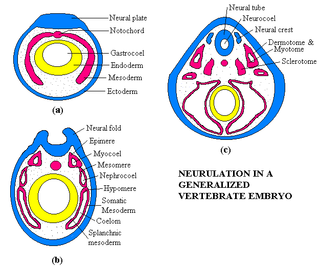
Coelum: the cavity surrounding the endodermal gut, formed by the hypomere. The coelom comes to be lined with splanchnic mesoderm axially and somatic mesoderm radially. The coelum is later divided into at least two cavities. The pericardial cavity contains the heart, and the pleuroperitoneal cavity contains the other internal organs. They are separated by the transverse septum.
Dermotome: The somites split into three segments, the most axial of which is the dermotome. This mesoderm is fated to become dermis.
Ectodermal placodes: ectodermal analogue of neural crest mesoderm and may be developmentally related. These are special populations of ectodermal cells that segregate during neurulation and sink into the body to form sensory receptors (visual, chemosensory etc. organs and related nerves) and as mesenchyme) cranial nerves.
Ectomesenchyme: same as mesenchyme, according to some sources. More strictly, mesenchyme derived solely from ectodermal cells of the neural crest.
Enterocoely: the (presumably primitive, but less common) mode of coelom formation illustrated in the figure in which the mesoderm enfolds the endoderm from the inception. The alternative mode is schizocoely.
Epimere: The original mesoderm differentiates into three sections. The epimeric mesoderm is the most dorsal region. It separates longitudinally into discrete clumps of mesoderm termed somites. Each somite is further split into dermotome, myotome, and sclerotome segments. The dermotome and myotome frequently remain connected for some time as in (c)) and are referred to as a dermomyotome. Epimeric mesoderm is ultimately fated to further differentiate into dermis, body muscles and vertebrae.
Gastrocoel: the space enclosed by the endodermal tube created by gastrulation.
Hypomere: The original mesoderm differentiates into three sections. The hypomeric mesoderm is the most ventral region. It is fated to further differentiate into limbs, peritoneum, gonads, heart, blood vessels and mesenteries.
Intermediate mesoderm: same as mesomere.
Lateral plate mesoderm: same as hypomere.
Mesenchyme: a sort of fourth germ layer formed by mixture of neural crest ectoderm and epimeric mesodermal cells. It is fated to differentiate into bone and dermis, among other structures.
Mesomere: The original mesoderm differentiates into three sections. The mesomere is the middle section. It is fated to further differentiate into the kidney and urogenital structures.
Myotome: The somites split, roughly axially, into three segments, the middle one of which is the myotome. This mesoderm is fated to become body musculature.
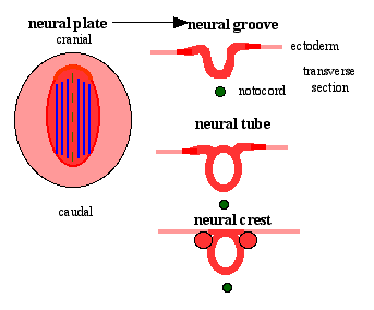
Neural crest: populations of mesodermal cells that separate from the neural tube as the neural tube pinches off from the surface ectoderm. It is fated to differentiate into visceral skeletal elements, nerve ganglia and, in combination with mesodermal cells, a variety of mesenchymal derivatives.
Neural folds: see figure (b)
 Neural Plate: dorsal thickening of ectoderm along the anterior-posterior axis. This invaginates and thickens to form the neural tube. Image from UNSW Embryology.
Neurocoel: the space enclosed by the neural tube.
Neurulation: the process of forming a dorsal ectodermal tube -- the neural tube.
Paraxial mesoderm: same as epimere.
Schizocoely: the (presumably derived, but more common) mode of coelom formation. It differs from the mode illustrated in the figure in that the mesoderm is originally the dorsal half of the invagination formed by gastrulation, rather than a separate layer which enfolds the endoderm. In schizocoel development, the presumptive mesoderm folds down over the endodermal tube and ultimately pinches off, so that, by the stage labeled (c), the two modes appear quite similar. The alternative mode is enterocoely.
Sclerotome: The somites split into three segments, the most medial of which is the sclerotome. This mesoderm is fated to become vertebral structures.
Somatic mesoderm: the outer wall of the hypomere. In association with ectoderm, it is fated to form limbs, gonads, and the peritoneum.
Somite: see epimere.
Splanchnic mesoderm: the inner wall of the hypomere. In association with endoderm, it differentiates into the heart and blood vessels, mesenteries, and (in mammals), extra-embryonic membranes.
checked ATW050108