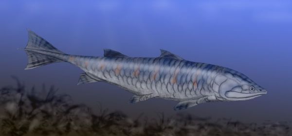
Grossius aragonensis, a sarcopterygian fish (family Onychodontidae) from the Middle Devonian of Spain, after Schultze (1973) and Janvier (1996),. Artwork by Nobu Tamura, 2008. Wikipedia, GNU Free Documentation/Creative Commons Attribution license.
| Sarcopterygii | ||
| The Vertebrates | More Basal Sarcopterygians |
| Vertebrates Home | Vertebrate | Vertebrate |
|
Abbreviated Dendrogram
Teleostomi
├─Neopterygii
└─Sarcopterygii
├─Psarolepis
└─┬─Achoania
└─┬─Grossius
└─┬─┬─Onychodontiformes
│ └─Actinistia
└─Rhipidistia
├─Dipnomorpha
└─┬─Rhizodontiformes
└─Osteolepiformes
├─Tristichopteridae
└─Elpistostegalia
├─Panderichthys
└─Tetrapoda
|
Contents
Overview |
 Grossius aragonensis, a sarcopterygian fish (family Onychodontidae) from the Middle Devonian of Spain, after Schultze (1973) and Janvier (1996),. Artwork by Nobu Tamura, 2008. Wikipedia, GNU Free Documentation/Creative Commons Attribution license. |
Psarolepis (continued from previous page)
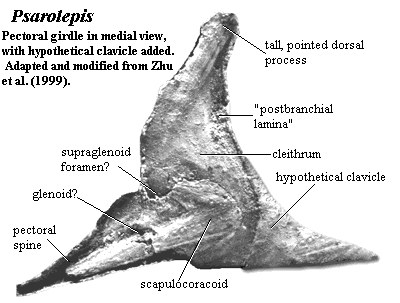 palate: upper jaw margin M-shaped in both anterior and palatal views [Y98]; premaxillae meet medially forming small part of anterior palate behind median rostral teeth [Y98]; large internasal cavities for parasymphysial tooth whorls [Z+99]; internasal cavities flanking dorsolateral margins of parasphenoid and extending posteriorly beyond it [Y98]; parasphenoid small & rhomboid [Y98]; parasphenoid with median longitudinal gutter containing hypophyseal canal, so that sides project ventrally beyond central region [Z98]; parasphenoid ascending process absent [Z98]; internal carotid artery does not penetrate parasphenoid [Y98];
lower jaw: in some (Silurian) specimens, left dentary overlaps right at the ventral part of the symphysis [ZS97]; postsymphysial pit for ligament to retract tooth whorls [L01]; dentary elongate with [at least?] 4 infradentaries [Z98]; three large foramina (as porolepiforms) mark boundaries between infradentaries [Y98] [Z+99]; infradentary fossae continued posteroventrally by furrow for articulation of submandibular [ZS97] [Z98]; possible splenial present on outer surface of mandible below symphysis [ZS97] (compare [Y98]: identifies this as an infradentary); lower jaw with at least 5 coronoids [Y98]; large medial dentigerous prearticular plate [Z98]; articular with kidney-shaped articular surface divided by ridge, probably for double-headed quadrate [Z98]; dentition: teeth with free pulp cavity [ZS97] (but see [Z+99]: polyplocodont dentition); median rostral toothed, with large fangs on tooth plate [Y98]; large parasymphysial tooth whorls [Z+99]; premaxilla and dentary with large inner teeth & irregular array of tiny outer teeth [ZS97] [Y98]; axial: probably with fin spines anterior to unpaired fins [Z+99]; appendicular: cleithrum, dorsal process tall, pointed [Z+99] [2]; very large pectoral spine extending from ridge between ventral and ascending lamina of cleithrum [Z+99] [1]; scapulocoracoid massive & plate-like [Z+99] other: large, closely-spaced pores on cosmine surface [Y98] [Z+99] pores are openings to pore-canal system [Z98]; enamel does not enter pores of pore canal system [ZS97].
Notes: [1] Z+99 point out the resemblance to placoderms and some acanthodians. [2] See image at left. [3] Y98 speculates that this groove served to anchor connective tissue.
Links: Sarcopterygii (Romer, 1955) Achoania gen. nov. Diagnosis. A ...; The Chinese Academy of Sciences; A new porolepiform-like fish, Psarolepis romeri; 2 (Oktober 1999)- Streiflichter; The Lineage of the Coelacanth - is there a link between ... ; Re: Nomina Conversa; Xiaobo YU's Kean University Home Page; Sarcopterygii;
References: Basden & Young 2001) [BY01], Long (2001) [L01], Miller et al. (2003) [M+03], Yu (1998) [Y98], Zhu & Schultze (1997) [ZS97], Zhu et al. 1999) [Z+99]. ATW040313.
 Achoania: A. jarvikii Zhu et al. 2001.
Range: Early Devonian (Lochkovian) of China (Yunnan).
Phylogeny: Sarcopterygii :: (Grossius + ((Onychodontiformes + Actinistia) + (Styloichthys + Rhipidistia))) + *.
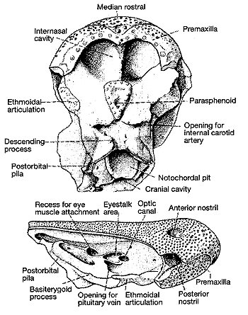
Characters: median rostral toothed, but does not separate premaxillae [Z+01]; premaxilla robust & rod-like, lacking posterodorsal process [Z+01] [1]; premaxilla does not reach orbit [Z+01] [1]; anterior nares open anteriorly (as Psarolepis) [Z+01]; posterior nares encased separate bone, presumably lacrimal [Z+01] [1]; intracranial joint angled anteroventrally [Z+01]; sphenethmoid with large descending processes anteroventral to notochordal pit [Z+01]; wide suborbital ledge [Z+01]; eyestalk present as heart-shaped area immediately posterior to optic foramen [Z+01]; posterodorsal to eyestalk is cup-shaped depression (myodome) supported ventrally by ridge [Z+01]; second depression ventral to eyestalk [Z+01]; postorbital pila robust, with posterodorsal branch flanking jugular foramen [Z+01]; internasal cavity large, occupying ~33% of ethmosphenoid floor [Z+01]; parasphenoid small, drop-shaped & flanked by flat rugose surface [Z+01]; carotids do not pierce parasphenoid [Z+01]; dermal bones covered by large-pore cosmine as in Psarolepis [Z+01]; 5 coronoids on lower jaw [ZY02].
Notes: [1] [ZY02] find that these are synapomorphies of the clade Achoania + crown group Sarcopterygii.
Image: (left) Sphenethmoid portion of skull in palatal and right lateral views, from [Z+01].
Links: Sarcopterygii (Romer, 1955) Achoania gen. nov. Diagnosis. A ... abstract of [ZY02]); TECH | Fossil fish in Chinese tale : A primitive sarcopterygian fish with an eyestalk..
References: Zhu & Yu (2002) [ZY02], Zhu et al. 2001) [Z+01]. ATW040320.
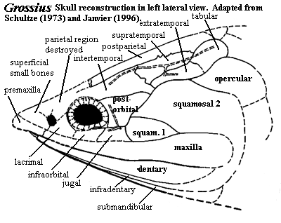
Grossius: G. aragonensis Schultze, 1973.
Range: Middle Devonian of Europe (Spain). There may be a problem with the date. In the original report, Schultze dates the only known material as early Middle Devonian (i.e. Eifelian) [S73]. However, in their review two decades later, Cloutier and Ahlberg state the age as Frasnian [CA96]. We have not been able to determine whether this is a typographical error or represents some re-assessment of the age of the "Moyuela Schichten."
Phylogeny: Sarcopterygii ::: ((Onychodontiformes + Actinistia) + (Styloichthys + Rhipidistia)) + *.
Characters: ~100 cm (but only skull known) [S73]; skull with pointed, three-cornered shape; only one pair external nares known (but anterior rostrum not recovered) [S73]; rostrum covered, by small bones [S73]; premaxilla present with dorsal process under cover of small bones [S73]; maxilla narrow, but becoming taller posteriorly [S73]; skull table formed mostly by elongate postparietals [S73]; intertemporal above orbit & postorbital, with posteroventral process reaching Squamosal 1 [S73]; intertemporal meets extratemporal & supratemporal posteriorly intertemporal above orbit & postorbital, with posteroventral process reaching Squamosal 1 [S73]; supratemporal with complex suture with postparietal [S73]; tabular posterior to extratamporal & supratemporal [S73]; orbits large & far from anterior end [S73]; lacrimal forming anterior orbital margin & probably posterior narial margin [S73]; estimate 20 plates in sclerotic ring [S73]; infraorbital separates lacrimal from jugal [S73]; jugal contacts orbit only at posteroventral corner [S73]; postorbital doubles in width dorsally [S73]; small squamosal 1 between maxilla & squamosal 2, reaching jugal and posteroventral edge of postorbital [S73] [1]; operculars not recovered except anterodorsal corner of (probable) opercular [S73]; palatoquadrate ascending process (nearly vertical posteriorly) visible in posterior orbit [S73]; behind orbit, palatoquadrate rises dorsally again to level of pterygoids with articulation to sphenethmoid [S73] [2]; sphenethmoid strongly ossified, surrounding cranial cavity & forming olfactory canals [S73]; ventral sphenethmoid in orbital region supports a deep parasphenoid [S73]; medial part of sphenethmoid & parasphenoid separate two deep hollows in the roof of the mouth for parasymphysial tooth spirals [S73]; more anteriorly, sphenethmoid cavities are deeper, separated only by wall of the sphenethmoid; [S73]; dentary thin, but becoming taller posteriorly [S73]; thin infradentaries and a submandibular ventral to dentary, with two overlapping gulars [S73]; large parasymphysial tooth whorls with 7-10 large main teeth, curving upward to level of center of orbit [S73]; tooth spirals also with small & medium-sized teeth (1 each?) [S73]; all parasymphysial teeth with striated enamel (40-50 lines/mm) on the lingual side [S73]; maxillary teeth lingual to ventral margin & hidden in lateral view [S73]; dentary teeth implanted in a gutter [S73]; plicidentine absent [S73]; hyomandibular with long anterior extension in dorsal recess of palatoquadrate [S73]; tooth-bearing plates probably present associated with branchial arches [S73]; vertebral elements (arch, pleuro- and intercentrum) found fairly far anterior, at level of most anterior portion of opercular [S73]; bones ornamented with shining tubercles, very closely spaced, appearing as a flat surface except under high magnification [S73]; scales unornamented except for single tubercle "peg" in area overlapped by other scales [S73]; scales smooth with clear pattern of concentric lines on inner surface [S73].
Notes: [1] squamosal 1 may be homologous to the quadratojugal. [2] See image of a very similar condition in porolepiforms. In both cases, the ascending process of the palatoquadrate is nearly vertical. The notch between the ascending process and the posterior body of the palatoquadrate apparently accommodates not only the ethmoid articulation of the palatoquadrate, but also provides room for the body of the m. levator arcus palatini.
References: Cloutier & Ahlberg (1996) [CA96], Schultze (1973) [S73]. ATW040328.
checked ATW041103