 The Standard Condition
| Bones: Dermal Bones | ||
| Vertebrates | Opercular Series: Overview |
| s ("Cladograms") |
||||||
| Vertebrates Home | Vertebrate | Vertebrate | Bones | Time |
|
Opercular Series
|
The opercular is a plate-like bone which covers the operculum, the structure which protects and operates the internal gills. There is simply no getting around the fact that, in order to have an opercular series of dermal bones, it is necessary to have an operculum. Consequently, the opercular series is present only in aquatic Osteichthyes. At this point, of course, some sniveling pedant will insist on bringing up the so-called operculum of the Holocephali. However, the likelihood that these soft tissue structures have any relationship whatsoever to the structures of interest to us, is beyond remote. Chondrichthyan gills operate in a quite different fashion. Accordingly, we will treat this disgraceful interruption with the scorn and derision it deserves. In fact, it is not even clear that the opercula of the Actinopterygii and Sarcopterygii are precisely the same structure, although we will assume that they are closely homologous.
The Standard ConditionThe Standard Condition (for a teleost) is shown at right. The opercular series comprises each gill cover (operculum) and generally consists of four bones. The preoperculars are flattened, J-shaped bones that usually bear heavy serrations on their posterior margins. It is not clear that they have the same origin as the operculum and the remainder of the series. Lying below each preopercular is a thin interopercular. Each interopercular normally possesses a small, dorsally-oriented projection on its anterior end. Posterior to each preopercular is the largest bone in the series, the opercular. Each somewhat triangular operculum has a large fossa on its upper anterior margin that articulates with the hyomandibular. Below each operculum is an oblong bone, the subopercular. The opercular series is connected to the lower jaw by a series of ligaments so that the jaws and opercula act in synchrony.
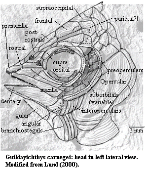
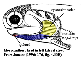 As we discussed in connection with the gulars, the opercular series seems to have developed from serial repetition of a simple overlapping laminar series of scales, as exemplified in some Acanthodii. The development from this stage is easily determined from the series at right and below, ultimately leading to the Standard Condition in teleosts. As noted above, it is not obvious that the interoperculars and -- in particular -- the preoperculars are derived in exactly the same fashion. In that connection, the very basal guildayichthyiform actinopterygian Guildayichthys demonstrates an interesting condition. Lund (2000). A moderately detailed image can be seen here. Guildayichthys has an extensive series of interoperculars parallel to the branchiostegal rays. They appear to have arisen by duplication of the entire branchiostegal series in a more dorsal position. This fish also has two long, thin preoperculars which, unlike the other members of the series, may well have been derived by the same sclerotic - orbital route which produced the jugal. However, that is a matter which can be taken up together with a more detailed consideration of those particular bones. The plot line is clear, even if we have not completely accounted for all of the characters.
 Some Functional History
Primitively, the operculum functioned as a sort of pump-and-valve-system for respiration. It seems likely that there has always been some connection between respiration and feeding in fishes. However, the operculum was essentially a respiratory structure, operating as part of a dual pump. The dual pump consists of a buccal and opercular pump, which act in synchrony. The first phase is he suction phase, in which the buccal and opercular cavities expand and water is drawn in. The opercular valve is closed at this point and lower pressure in the opercular cavity draws water across he gills. In the second, force phase, the mouth is closed and he opercular valve open. Muscle compression forces water posteriorly and out. The result is unidirectional flow. respiration.pdf.
At the level of the Halecostomi and above, things get more involved as the nature of the jaw changes. In essence, the posterior end of the lower jaw is depressed -- dropping the floor of the mouth -- by using the interopercular as an extension of the lower jaw. The trick is done by contraction of the m. levator operculi (derived from the primitive opercular adductor muscle). The levator causes a dorsal rotation of the entire opercular series (operculum, subopercular and interopercular) which is applied as a posteroventrally directed force on the posterior end of the jaw via the interoperculomandibular ligament. Lecture 2.
Wakarimashta?
Let's try it again: review the image of Amia at right. if one could grip the opercular series like a dial and twist it counter-clockwise, that would force the far end of the interopercular down and to the right. The left end of the interopercular and the right end of the lower jaw are connected by a ligament, so that end of the jaw moves the same way. That drops the whole floor of the mouth.
Now, if we're an advanced teleost, the whole thing looks like this:
1. First the fish must prepare. During the preparatory phase the volume of the buccal cavity is reduced. Basically the floor and the sides (i.e. the hyoid region and the suspensorium region) of the fish's mouth squeeze together.
2. Next the fish expands the volume of the buccal cavity, opens its jaws, and in some species also protrudes its upper jaw. During this phase water and prey are sucked into the fish's mouth. There are three musculoskeletal couplings that are involved in this expansion phase.
a. epaxial muscles lift the cranium and the roof of the mouth, expanding the buccal cavity.
b. The lower jaw is depressed. This occurs when the operculum is rotated via the levator operculi and interopercular-mandibular ligament as explained above.
c. Additionally, the hypaxial muscles depress the lower jaw. Hypaxial and the sternohyoid muscles act on the bones of the floor of the mouth (the hyoids) and also depress the lower jaw.
3. During the compression phase, the jaws are closed by the adductor mandibulae (see Fig. B below) Also the sides of the fishes mouth are squeezed in (the suspensorium is adducted), and the cranium returns to its original position. If the buccal cavity is being squeezed in, where does the water in the mouth go? During the compression phase, the operculum valve opens and the water in the buccal cavity flows over the gills, past the operculum and out of the fish. The protruded jaw returns to its original position also.
4. During the recovery phase, the muscles and bones return to their original positions. The length of this phase is longer when large prey are consumed. Perch supplement2.pdf.
These complex steps are coordinated with movements of the gills and the strange dance of maxilla and premaxilla which seems almost backward to those of us more accustomed to the simple-minded hinge of the tetrapod jaw. See, e.g., Premaxilla.
Links: Lecture 2 (important stuff on helecostomes); Perch supplement2.pdf; Untitled Document (important figure & figures of mechanism above); MsoDockBottom; gibbpage/pvfeed.pdf (the case of flatfish); respiration.pdf; Biology 356; The materials provided with the previous exam; Fall'96Syllabus. ATW020817.
checked ATW050705