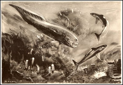
 The early shark Cladoselache flees from the arthrodire superpreditor Dunkleostes. Artwork by Zdenek Burian, in Joseph Augusta, Prehistoric Animals (Paul Hamlyn Ltd)
| Chondrichthyes | ||
| The Vertebrates | The Crown Group |
| Vertebrates Home | Vertebrate | Vertebrate |
|
Abbreviated Dendrogram
Cephalaspidomorphi
├─Placodermi
│
└─Eugnathostomata
├─Chondrichthyes
│ ├─┬─Eugeneodontida
│ │ └─Chondrichthyes (Crown)
│ ├─┬─Cladoselachida
│ │ └─Symmoriida
│ │ ├─Symmoriidae
│ │ └─Holocephali
│ └─Elasmobranchii
│ ├─Xenacanthida
│ └─Ctenacanthiformes
│ ├─Ctenacanthidae
│ └─Euselachii
│ ├─Hybodontiformes
│ └─┬─Synechodontiformes
│ └─Neoselachii
└─Teleostomi
|
Contents
Overview |
 The early shark Cladoselache flees from the arthrodire superpreditor Dunkleostes. Artwork by Zdenek Burian, in Joseph Augusta, Prehistoric Animals (Paul Hamlyn Ltd) The early shark Cladoselache flees from the arthrodire superpreditor Dunkleostes. Artwork by Zdenek Burian, in Joseph Augusta, Prehistoric Animals (Paul Hamlyn Ltd) |
 The Crown Group and the Cladoselachida
This is the chondrichthyan crown group, defined as ratfish + rays. On one side we have odd symmoriids, strange stethacanthids, and downright bizarre holocephalans, including the extant chimaeriforms. On the other side, we find the elasmobranchs, a conservative and dignified lot consisting of the living sharks and rays (Neoselachii) and their equally stuffy and conservative ancestors. Somewhere near the intersection of these two incompatible groups, trying desperately to hold the family together, are the Cladoselachida.
Despite the name, the Cladoselachida may not be sharks, as the word is normally used, and are certainly not a clade of cladodont sharks. It isn't clear what it takes to be a "cladodont." The word is sometimes used to denote one of four vague grades of elasmobranch: cladodont, xenacanth, ctenacanth, and hybodont. It also describes a type of chondrichthyan tooth morphology which has no useful phylogenetic implications. In fact, the group is named for a genus, Cladodus, which is a nomen nudum, "a form genus sustained only by our ignorance of skeletal remains unequivocally related to the rather diverse tooth form known as 'cladodont.'" Schaeffer 1981); Maisey 2001a) (quoting Schaeffer).
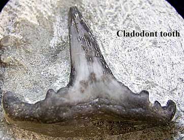
For what it may be worth, the typical cladodont tooth, as shown in the image, features a large, relatively skinny main cusp flanked by 2-4, roughly symmetrical subsidiary cusps on each side. The outermost cusp is generally the largest of the subsidiary cusps. The main cusp, in particular, bears longitudinal striations. All cusps bulge slightly on the buccal side, leaving a broad lingual shelf. Williams 1998). The shelf overlies a bulge of attachment bone referred to as a "torus." Maisey (1977, 1983). However, this type of dentition is not restricted to the classical Cladoselachida. In fact, they may be more typical of ctenacanths (whatever this means -- see Ctenacanthidae). Nor do these teeth diagnose any reasonably defined clade of chondrichthyans. In fact, now that we think of it, we've seen conodont elements which could be described in practically the same terms.
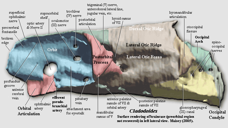
Consequently, there's not a great deal we can say about Cladoselachida or Cladodus. However, we have entirely too much to say about one particular putative cladoselachian, Cladodoides. Cladodoides is one of two isolated braincases recovered from the Late Devonian of Wildungen, Germany. Both braincases were found stratigraphically and geographically near some jaw material on which Jaeckel (1921) erected the genus Cladodus and the species wildungensis. Both braincases were originally referred to C. wildungensis, although there was no diagnostic basis for doing so. In 1937, the two braincases were separately described by Eric Stensiö and Walter Gross. Gross confused things further by referring Stensiö'sspecimen to a new species, C. hassiacus. There matters stood for about 60 years, until Dr. John Maisey of the American Museum of Natural History decided to have another look at this famous braincase (Gross's specimen). In view of the uncertain affinities of the specimen and the vagueness of Cladodus, Maisey redesignated Gross's specimen Cladodoides. Maisey (2001a). A full redescription followed in Maisey 2005).
 By way of background, Maisey started producing a series of remarkable shark brain papers in about 1999 -- a series which continues to this day.Something obviously happened which sparked a sudden and pronounced intensity in Maisey's work. The closest analogy we can recall at the moment is Kobe Bryant of the Los Angeles Lakers. Bryant was always one of the NBA's best supporting players, with solid, consistent play and strong defense. Then, quite suddenly, in mid-career, Bryant went from being a top supporting player to producing legendary individual performances, such as his 81 points against Toronto in January, 2006. True, Dr. Maisey will probably never score 81 points against Toronto; but, then again, Kobe Bryant is probably quite weak on Paleozoic shark anatomy.
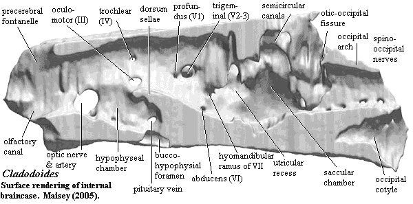
But tiresome sports analogies aside, Maisey (2005) is a classic paper for two reasons. First, it touches so many of the big issues in early vertebrate paleontology.So, for example, it speaks in some detail about some recent startling changes in the developmental biology of the jaw (Cerny et al., 2004), the relationship between the orbital articulation of sharks and the basicranial articulation of tetrapods, the relationship between the extrinsic eye muscles in the basal gnathostome groups, the comparative anatomy of the semicircular canals in chondrichthyans and other early vertebrates, the origins of the metotic fissure -- in fact, just about all of the things that make early vertebrate paleontology interesting. Second, it provides a wonderful example of what technology can do for paleontology. Maisey not only made extensive use of the computerized tomography facilities at the University of Texas, but went on to process the CT data with additional software which made it possible for him to visualize internal "surfaces" in three dimensions. For example, the image shows the internal surface of the braincase, as if the specimen had been cut in two and prepared on the inside. Actually the braincase is intact and the inner surface is both invisible and covered with matrix.
Best of all, the paper is free for the downloading at the AMNH website. This is particularly fortunate, since it provides us with an iron-clad excuse not to attempt the impossible job of summarizing the entire paper. Instead, we will take on the more congenial (if equally lengthy) task of wandering down just one of the legion of scenic anatomical trails which radiate from the braincase of Cladodoides. Specifically, we will look at a vessel bearing the following preposterous name:
The What?
As usual, we must begin with a careful study of nomenclature, using well-established rules of anatomical deconstruction. In approaching any new item of cranial anatomy, we should first apply Przynowitzkyinskya-Krzybblyskoumi's Rule: the complexity of an anatomical label is inversely proportional to the size and importance of the object to which the label adheres, unless the label includes the name of some guy who might possibly be Italian. So, the EPA is clearly a small feature, perhaps of limited functional significance. Second, we make use of a lemma of Law of Maximum Perversity, namely: all recognizable roots in an anatomical name will be misleading. Since we recognize the root terms branchial (relating to gills) and artery a vessel which supplies blood from the heart) we can be reasonably certain that the EPA has nothing to do with gills and is probably a vein. Finally, we will add that there exists a fairly broad literature on the structure of the EPA. This should suggest the relevance of the Canon of Reciprocal Befuddlement, an analogue of the famed Uncertainty Principle. The Canon holds that: for each anatomical structure, there exists a point (п ) after which any increased knowledge of structure proportionately decreases our understanding of how the hell the thing can possibly perform its supposed function -- and vice-versa.
Combining these rules, we may infer that the efferent pseudobranchial artery i) is a relatively small and/or unimportant vessel which (ii) is probably a vein with no obvious connection to gills, and (iii) performs a function which is not well understood. These conclusions are largely negative, but -- as we will see -- fundamentally sound.
Range: fr lwD.
Chondrichthyes : Squatinactida + Orodontida + (Eugeneodontida + Petalodontiformes) + * : (Cladoselachida + Symmoriida) + Iniopterygii + Elasmobranchii.
Pelvic clasper.
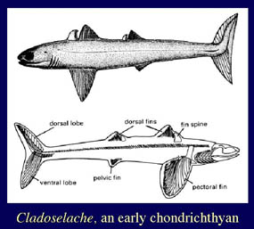 Cladoselachida: "Cladodus," Cladodoides, Cladoselache.
Range: From the Early Devonian. Last appearance depends on what is included and whether the group is paraphyletic. Certainly to the Late Devonian under any definition.
Phylogeny: Condrichthyes Crown) : Symmoriida + *. Cladodus is a nomen nudum. If we give this group a reasonable phylogenetic definition, say Cladoselache > Cobelodus, it may be synonymous with Crown Group Chondrichthyes, or an even more inclusive clade.I
Characters: Mouth terminal; teeth cladodont (3-pointed); palatoquadrate firmly connected to braincase; braincase short, with long post-orbital process; 5 gill slits; notochord unrestricted; 2 dorsal fins; short spine anterior to 1st (& 2nd?) dorsal fin; fin margins supported by ceratotrichia; paired fins also contain unjointed radials articulating directly on girdle or metapterygium; fin spines lack enameloid (c.f. enameloid ontogeny) (all dentine); no anal fin; tail large & lunate, with upper chordal) lobe reenforced by cartilagenous plates; no pelvic claspers; few scales; scales only on fin margins and around orbit. Perhaps similar to modern fast predators (e.g. tuna); known to have fed on actinopterygians & perhaps conodonts.
Geol 437 sharks, Fall 1995; 70d10.pdf German).
ATW060324.
checked ATW060126