Rhizaria
Eukarya Rhizaria

Rhizaria


Abbreviated Dendrogram
Eukarya ├─Metamonada └─┬─Discicristata └─┬─Rhizaria │ ├─Radiolaria │ │ ├─Acantharia │ │ └─Polycystinea │ └─┬─┬─Acetosporea │ │ └─┬─Xenophyophorea │ │ └─Foraminifera │ └─┬─Phytomyxea │ └─Cercozoa └─┬─┬─Chromalveolata │ │ ├─Alveolata │ │ └─Chromista │ └─Plantae └─Stem Metazoa ├─Fungi └─Metazoa
Contents
Introduction
Phylogeny
Diversity
   Radiolaria
   Acantharia
   Polycystinea
   Acetosporea
   Foraminifera
   Phytomyxea
   Cercozoa
References

Cross Reference Palaeos.org


Introduction

Unidentified radiolarianThe clade Rhizaria of unicellular eukaryotes was named very recently (Cavalier-Smith, 2002), but has rapidly ingratiated itself as an industry standard. It contains a large number of mostly amoeboid organisms, including such significant groups as the radiolarians and foraminiferans.

So far, Rhizaria seems to be supported solely by molecular data – there are no morphological characters unique to the clade. Most are biciliate amoeboflagellates, at least at some point in the life cycle – though many have dispensed with flagella altogether. Pseudopodia are root-like reticulopodia, filopodia and/or axopodia – not broad lobopodia as in Amoeba. All of these features can, however, be found in members of other clades. Nevertheless, the Rhizaria are supported by both rRNA and actin trees Cavalier-Smith & Chao, 2003; Nikolaev et al. 2004), and are probably here to stay.

Image: unidentified radiolarian from the Hamilton College Electron Microscopy site.

© Christopher Taylor 2004. CT041217

Rhizariate Phylogeny

Rhizaria ├─Radiolaria │ ├─Polycystinea │ └─┬─Sticholonche │ └─Acantharea └─┬─┬─Gromiidae │ └─┬─Ascetosporea │ └─Foraminifera └─┬─Phytomyxea └─┬─Desmothoracida └─'core Cercozoa' (including Phaeodarea)

SSU rRNA and actin trees both give a similar picture of rhizarian phylogeny – the phylogeny below is derived from Nikolaev et al. (2004) and Polet et al. (2004). The positions of Sticholonche and Ascetosporea, however, should be treated with some suspicion.

Cavalier-Smith (2002; Cavalier-Smith & Chao, 2003) previously suggested a monophyletic clade, Retaria, formed by Radiolaria and Foraminifera, characterised by reticulose pseudopods in both groups, and supported weakly by molecular phylogenies. More recent analyses fail to support this grouping, and reticulopodia probably evolved independently in the two groups – an adaptation to large size in both? Instead, Foraminifera group with Gromia, a marine amoeboid with smooth filopodia that produces an organic test like many basal Foraminifera (Longet et al. 2003).

© Christopher Taylor 2004. CT041217

Rhizariate Diversity

The Rhizaria can be thought of as being composed of Radiolaria, Foraminifera, and Cercozoa. That's fortunate, since we are going to treat Rhizaria in that very manner.

Radiolaria

RadiolariaLarge, planktonic forms that produce a glassy, intricate shell. A protein capsule divides the cytoplasm into inner and outer compartments. The capsule is perforated by numerous scattered pores through which the axopodia pass. All radiolarians secrete strontium sulphate at some point in the life cycle – as the adult shell in Acantharea, and as crystals in 'swarmer cells' produced during asexual reproduction in Polycystinea. The adult shell in Polycystinea is siliceous. Axopodia joined by cross-branches. Endosymbiotic algae are usually present (Polet et al. 2004).

The name 'Radiolaria' has a particularly ghastly history – traditionally, it has included three glassy-shelled taxa, the Polycystinea, Acantharea and Phaeodarea. The monophyly of these three groups has long been suspect, and Radiolaria has been used for Polycystinea and Acantharea excluding Phaeodarea, Polycystinea and Phaeodarea excluding Acantharea, and Polycystinea alone. Phaeodarea are not closely related to the other two taxa (see below), but Acantharea and Polycystinea form a monophyletic group (Nikolaev et al., 2004; Polet et al., 2004). In the absence of a better name, we elected have to keep using Radiolaria for a mere segment of its previous self. It may be arbitrarily defined as organisms closer to Thalassicola (Polycystinea) than to Allogromia (Foraminifera) or Cercomonas (Cercozoa).

Radiolaria have also been included in the past as part of a taxon Actinopoda along with a number of radial axopod-bearing organisms called Heliozoa. 'Heliozoa' has since turned out to be a rampantly polyphyletic group – examples have been reclassified as chromists and opisthokonts (Mikrjukov, 2000; Cavalier-Smith & Chao, 2003; Nikolaev et al., 2004). One past heliozoan, Sticholonche, was found by Nikolaev et al. (2004) to cluster with Acantharea, but support values were low, and this seems suspicious. Its inclusion in this position would, for instance, imply that the intracellular capsule either evolved independently in the Acantharea and Polycystinea, or that it was lost in the ancestor of Sticholonche. Sticholonche is most notable for the way that the axopods are actually used to actively row the organism through the water (Febvre-Chevalier, 1990).

Polycystinea have an extensive fossil record back to the late Precambrian (Cachon et al. 1990), and are very important in biostratigraphy.

© Christopher Taylor 2004. CT041218

 Radiolarians

Polycystinea

The Polycystinea (sometimes spelled Polycistinea or Polycystina) are one group of the Radiolaria. These are not just "small shelly fauna," they are tiny shelly fauna made up of single, if rather complex, cells. The shell turns out to be made of amorphous silica -- essentially sand -- without the admixture of organics that characterize similar forms. Polycystinea are exclusively marine but found in great numbers in the oceans. Their fossil record goes back almost a billion years, well into Precambrian time.

Like other radiolarians, the cytoplasm of Polycystinea is divided into ectoplasm and endoplasm by a perforated protein capsule -- not the nuclear membrane, but a novel structure unique to this group. The endoplasm forms a central medulla enclosed by this  porous, membranous capsule. The nucleus is inside this central region. The ectoplasm is outside the capsule and forms the region known as the cortex (or calymma). The visible remains shown in the image are made up of perforated tests (the "shells"). In life, these are located in the ectoplasm. Polycystinates extend pseudopods supported by a complex microtubular array (axopods) which originate in the endoplasm. The pseudopods pass through pores in the test and extend, covered with a thin layer of cytoplasm, from the surface of the cell. Spines of the test, if any, also pass through the capsule and extend, covered with cytoplasm, from the surface of the cell. The ectoplasm is often vacuolated and frequently contains photosynthetic zooxanthellae.

The endoplasm actually contains all of the organelles normally associated with a "normal" heterotrophic eukaryotic cell, including mitochondria, a nucleus, and a cytoskeleton. The ectoplasm is largely filled with digestive vacuoles, symbiotic algae, and the test. From an evolutionary standpoint, the Polycystina appear to be one step towards a whole different type of biological organization based on a 3-compartment cell, rather than the 2-compartment cell of metazoans. In fact, a number of polycystinean species are colonial. It is interesting to speculate on what might have evolved on this model, had circumstances been different.

ATW030819. Text public domain. No rights reserved.

Acantharea

The Acantharea already have their own page.

The foraminiferan clades are highly contentious, which is too bad. The evolution and diversity of these sturdy, testate Eukarya form an important part of Mesozoic history and stratigraphy. A better understanding of their ?Cambrian beginnings might give us a bater handle on their later development. We treat only one group of stem Foraminifera here, the Acetosporea.

Acetosporea

Haplosporidium

Amoeboid, non-flagellate parasites of shellfish, comprising the orders Haplosporida and Paramyxida. Cavalier-Smith & Chao (2003) found weak support for an association with the plant-parasitic Phytomyxea and included both in a subphylum Endomyxa. Nikolaev et al. (2004) found Haplosporida as the closest relatives to Foraminifera. Historically, they have been regarded as similar to Microsporidia. Watch this space. Haplosporidium, Urosporidium, Marteilia.

Image:  Haplosporidia from the Leech Lab site of Dr. Mark Siddall at the AMNH.

 Foraminifera

Foraminifera

Amoeboid organisms characterised by reticulate, granular pseudopodia (hence the often-seen alternative name Granuloreticulosa). Mostly marine; endosymbiotic algae often present. The majority of Foraminifera produce a test of some form or other – mostly calcareous, but agglutinated or organic in more basal forms. One group of basal agglutinated-test Foraminifera became sessile, and a subgroup of this line took to growing to Brobdingnagian proportions – the Xenophyophorea. Pawlowski et al. (2003).

Foraminifera, especially the calcareous forms, have a fossil record stretching back to the Cambrian (Lee, 1990), and are especially important biostratigraphically.

The Xenophyophorea are either Foraminifera, or possibly the sister group of Foraminifera. These bizarre, gigantic protists are commonly several centimeters in diameter and are discussed on their own page.

Text © Christopher Taylor 2004. CT041218

More on the Foraminifera

Finally, the cercozoan group:

Phytomyxea

Plasmodial plant parasites, primarily known for the problematic Plasmodiophora, the cause of club root in brassicas. Appear to be the most basal branch of Cercozoa. Generally regarded in the past as fungi of some sort, and so referred to as Plasmodiophoromycota or some variation thereof. Phytomyxea at least has the virtue of being a much shorter name.

Text © Christopher Taylor 2004. CT041218

 Cercozoa

Cercozoa, originally named by Cavalier-Smith in 1998, is a diverse group of taxa united solely on molecular grounds, but supported by a number of genes (Longet et al., 2003). As generally circumscribed, Cercozoa also includes Gromiidae and Phytomyxea, but these more divergent taxa have been listed separately here to show their relative phylogenetic positions (and also to avoid having to lump Foraminifera in with the Cercozoa). For a brief period before 1998, the clade soon to be called Cercozoa was referred to as Rhizopoda, as it included a large proportion of the species previously included in that form-taxon (specifically those bearing filose pseudopodia). But as many rhizopods were not in this group, including the best-known example, Amoeba, and many Cercozoa are flagellates rather than amoeboid, the name Cercozoa is much more welcome.

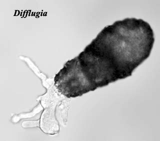
Amongst notable members of the Cercozoa are amoeboid forms such as Difflugia, which produce agglutinated tests that may be fossilised (the record extends back to the Neoproterozoic Finlay et al., 2004), and the Chlorarachnea (e.g. Chlorarachnion), marine amoeboid organisms which possess chloroplasts derived from a secondary endosymbiosis with a green alga. Cavalier-Smith, 2003). The nucleus of the endosymbiont in Chlorarachnion, in fact, has not fully degraded as in most secondarily plastid-bearing eukaryotes, and the chloroplast retains a small nucleomorph contained within the surrounding membranes.

Nikolaev et al. (2004) and Polet et al. (2004) both found Phaeodarea to also be nested within Cercozoa, though a strong association with any particular taxon or taxa was not supported. Phaeodarea were traditionally included in Radiolaria, and share with Acantharea and Polycystinea the traits of a glassy shell (formed of a combination of silica and organic material in Phaeodarea) and a capsule dividing the cytoplasm into inner and outer compartments. In the Radiolaria as here defined, however, the capsule is thin and perforated by numerous pores – in Phaeodarea, the capsule is much thicker, and usually only three pores pass through it, the astropylum and and usually two parapyla situated at the opposite pole. The astropylum forms a cone-like cytopharynx that is used for the ingestion of food items. Phaeodarea also bear a phaeodium, consisting of balls of darkly pigmented waste matter, usually near the astropylum. Phaeodarea also lack algal endosymbionts and cross-branches between the axopods. Polet et al. (2004).

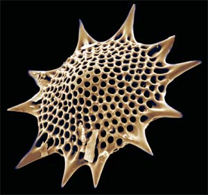
Image: Difflugia from the Droplet site.

Text © Christopher Taylor 2004. CT041218


checked ATW061220, edited RFVS111204