| Palaeos: |  |
Glossary |
| EUKARYA | Glossary A-b |
| Page Back | Unit Back | Unit Home | References | Glossary | Pieces |
| Page Next | Unit Next | Life | Dendrogram | Taxon Index | Time |
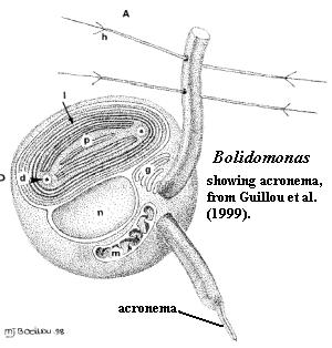
 Acronema:
a short, thin terminal extension at the end of a flagellum. Image from Guillou
et al. (1999).
Actin: the most abundant single protein in most eukaryotic cells. Microfilaments are essentially actin polymers. Actin may exist as a globular monomer (G-actin) or as a linear polymer (F-actin). See links at Microfilament Links.
Alpha chitin: See chitin.
Alveoli: An important synapomorphy of the Alveolata. The alveoli normally appear as small vesicles in or under the plasma membrane. They are not associated with ribosomes and do not have any detectable contents. They do not seem to be connected with any membrane system other than the plasma membrane. It is now believed that the alveoli are part of a complete second inner membrane system.
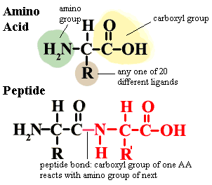
 Amino
acid: the fundamental building block of proteins. There are twenty
different amino acids normally found in proteins. All have the general
structure shown in the figure. In proteins, the amino acids are joined by
peptide bonds as shown in the image. Notice that the central carbon atom
has four different ligands. It is therefore asymmetrical and can exist in
two mirror image forms (enantiomers), known as L
and D enantiomers.
Proteins in living organisms are all made from L-amino
acids. However bacterial cell walls and a few other structures
incorporate some D-amino
acids. A few naturally occurring amino acids are not normally found in
proteins and are not specified in the genetic code.
Ornithine (R = (CH2)3NH2) is one example.
These non-protein amino acids are common intermediates in a variety of
metabolic pathways. Finally, some amino acids may be chemically modified
after they have been incorporated into proteins.
Antapical: posterior.
Antenna pigment: a light-absorbing pigment which is used to transmit light energy by absorbing at one wavelength and emitting at another. In the usual case, light is initially absorbed by a pigment which is sensitive to a range of wavelengths outside the range which chlorophyll can absorb. Antenna pigments act as transformers to modulate the wavelength so that the light energy can be passed to chlorophyll and used in photosynthesis.
Apical: anterior.
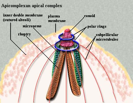
 Apical
complex: the characteristic organ complex of the Apicomplexa,
including rhoptries, micronemes,
polar rings, and, if present, the conoid.
Autogamy: "fertilization" between two daughter gametes of the same gametocyte. Typically this occurs without complete separation. That is, the nucleus of the diploid or tetraploid gametocyte divides without DNA synthesis and without complete cytokinesis (complete separation of daughter cells). The resulting cell then behaves like a zygote resulting from fertilization.
Axoneme: the fundamental 9+2 doublet microtubule structure at the core of the eukaryotic flagellum. The axoneme arises from the basal body and inserts into the axosome.
Axopod: Thin processes (a few microns in diameter but up to 500m long), supported by complex arrays of microtubules, that radiate from the bodies of radiolarians and various other cells. Each axopod is composed of a core of microtubules, the axial rod, which arises in the medulla, and a thin covering of cytoplasm enclosed in the cell membrane. An axopod which comes in contact with a food item quickly retracts, and the item is phagocytosed.
Axosome: the thin extension of the plasma membrane and associated cytoplasm that covers the flagellum. The microtubule doublets originating in the basal body insert into the axosome. See image at flagellum.
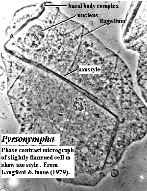
 Axostyle:
The microtubule-containing organelle known as the axostyle found in certain
zooflagellates propagates undulatory bending waves similar to a flagellum or
cilium. Electron microscopy studies show that the motile axostyle in the
wood roach commensal oxymonad Saccinobaculus and the termite protozoan Pyrsonympha
contains several thousand singlet microtubules interconnected by cross-
bridges. The microtubules are organized into rows, and the microtubules
within the rows are connected to each other by regularly occurring linkers or
intra-row bridges. In turn, the rows of microtubules are interconnected by less
regularly occurring cross-bridges or inter-row bridges. The intra-row bridges
appear periodic along the tubules with a spacing of 16 nm. The inter-row
bridges are not strictly periodic and can be oriented at varying angles to the
axis of the microtubule. Langford
& Inoue (1979). This reference also contains several good electron
micrograph images illustrating the ultrastructure described above. The
oxymonad Saccinobaculus
has an especially impressive axostyle, with many images on the
web.
Biflagellate: having two flagella.
Blepharoplast: same as basal body.
| Page Back | Page Top | Unit Home | Page Next |