| Palaeos: | Bones: Dermal Bones | |
| Vertebrates | Orbital Series: Jugal (2) |
| Page Back | Unit Back | Unit Home | Unit References | Unit Dendrograms ("Cladograms") |
Glossary | Taxon Index |
| Page Next | Unit Next | Vertebrates Home | Vertebrate References | Vertebrate Dendrograms | Bones | Time |
|
Bones
|
Dermal Bones
Facial Series |
Orbital Series
|
 Ornithischia:
The Ornithischia frequently have lateral elaborations of the jugal, particularly
the posterior process, bearing a variety of bosses, knobs, horns and other
ornaments. This sort of ornament seems to have developed a number of
times: in the Ankylosauria
(Coombs & Maryanska, 1990), Heterodontosauridae,
and Ceratopsia.
However, this is probably a common consequence of herbivory, since similar
developments are seen in pareiasaurs, Estemmenosuchus,
and (less clearly) in rhynchosaurs.
The lateral expanse of the jugal is simply too good a place to hang cheeks and
jowls. The development in the Hadrosauroidea
has more phylogenetic and anatomical interest. Here, the jugal is rather
solidly attached at both ends, i.e., to the quadratojugal and the
maxilla. However, the bone is thin, expanded dorsoventrally, and has only
slight or sliding contact with the lacrimal and postorbital. Head
(1998); Sereno (1986), vide Norman
(1990). Presumably, this permitted the jugal to bow outward to accommodate
the unique hadrosaur style of chewing. In more derived forms,
the jugal reacquires its accustomed scarf joint with the quadratojugal and loses
contact with the ectopterygoid, permitting even greater flexibility. Head
(1998).
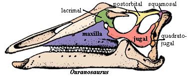
Ornithischia:
The Ornithischia frequently have lateral elaborations of the jugal, particularly
the posterior process, bearing a variety of bosses, knobs, horns and other
ornaments. This sort of ornament seems to have developed a number of
times: in the Ankylosauria
(Coombs & Maryanska, 1990), Heterodontosauridae,
and Ceratopsia.
However, this is probably a common consequence of herbivory, since similar
developments are seen in pareiasaurs, Estemmenosuchus,
and (less clearly) in rhynchosaurs.
The lateral expanse of the jugal is simply too good a place to hang cheeks and
jowls. The development in the Hadrosauroidea
has more phylogenetic and anatomical interest. Here, the jugal is rather
solidly attached at both ends, i.e., to the quadratojugal and the
maxilla. However, the bone is thin, expanded dorsoventrally, and has only
slight or sliding contact with the lacrimal and postorbital. Head
(1998); Sereno (1986), vide Norman
(1990). Presumably, this permitted the jugal to bow outward to accommodate
the unique hadrosaur style of chewing. In more derived forms,
the jugal reacquires its accustomed scarf joint with the quadratojugal and loses
contact with the ectopterygoid, permitting even greater flexibility. Head
(1998).
 Saurischia:
The Saurischia are characterized by a synapomorphy of the jugal. The posterior process is forked and grasps the quadratojugal with two or three
tines. See, e.g., figure and discussion at Sinraptoridae;
Currie & Zhao (1993).
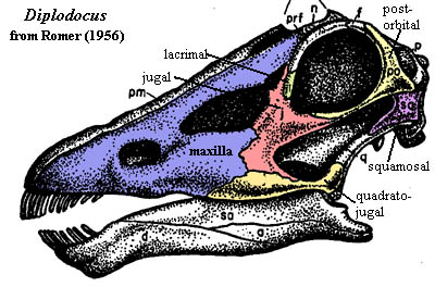
Another peculiar jugal worth noting is that of the Neosauropoda.
The retraction of the nares to the top of the skull causes a bizarre
rearrangement with the result that the "anterior" and
"posterior" processes of the jugal may actually merge, as shown in the
image of Diplodocus.
For those who may not have had much practice with sauropod skulls,
remember that the cavity in the anterior part of the maxilla is not a
nostril. It is a second antorbital fenestra. The nostrils are on top
of the head, above the orbits.
Saurischia:
The Saurischia are characterized by a synapomorphy of the jugal. The posterior process is forked and grasps the quadratojugal with two or three
tines. See, e.g., figure and discussion at Sinraptoridae;
Currie & Zhao (1993).
Another peculiar jugal worth noting is that of the Neosauropoda.
The retraction of the nares to the top of the skull causes a bizarre
rearrangement with the result that the "anterior" and
"posterior" processes of the jugal may actually merge, as shown in the
image of Diplodocus.
For those who may not have had much practice with sauropod skulls,
remember that the cavity in the anterior part of the maxilla is not a
nostril. It is a second antorbital fenestra. The nostrils are on top
of the head, above the orbits.
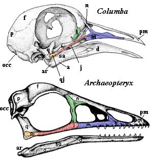 Aves: Oddly enough, one of the first adaptations of birds was a fairly
radical reorganization of the jugal area and the reduction of the jugal to a
slim jugal
bar, actually consisting of a fused jugal and quadratojugal, connecting
the maxilla and upper beak to a quadrate which is moveable on the occiput.
This development was presumably preceded by a loss of the postorbital bar since,
even in Archaeopteryx, the postorbital is present -- at most -- as a thin
ridge on the anterior face of the quadrate. A slight contact with the
lacrimal is maintained, but there is no palatal exposure. Note that,
despite the reorganization, the jugal maintains all of its primary articulations
except for the postorbital, and even this is a close call.
Aves: Oddly enough, one of the first adaptations of birds was a fairly
radical reorganization of the jugal area and the reduction of the jugal to a
slim jugal
bar, actually consisting of a fused jugal and quadratojugal, connecting
the maxilla and upper beak to a quadrate which is moveable on the occiput.
This development was presumably preceded by a loss of the postorbital bar since,
even in Archaeopteryx, the postorbital is present -- at most -- as a thin
ridge on the anterior face of the quadrate. A slight contact with the
lacrimal is maintained, but there is no palatal exposure. Note that,
despite the reorganization, the jugal maintains all of its primary articulations
except for the postorbital, and even this is a close call.
Synapsida: Recall that in the Standard Condition, the jugal contacts the squamosal. In the Diapsida, this connection is broken by the appearance of the lower temporal fenestra. In synapsids, the connection is generally maintained, with profound results. We say "generally," since the connection is believed to exist in the Eothyrididae, but is not known with complete certainty. Langston (1965). In some of the Varanopseidae, the squamosal definitely does not contact the jugal. Reisz et al. (1998); Romer & Price (1940). However, the subtemporal bar is slender and fragile in all of these forms, and we may maintain a healthy degree of uncertainty without intending any criticism of these authors.
 Therapsida:
Certainly, by the level of the therapsids, the jugal-squamosal connection is
well-established. See the images at the Profusely
Illustrated Guide. In fact, the squamosal seems to have developed by a
sort of competitive exclusion of the quadratojugal, which eventually disappears
entirely, with all of its functions being gradually subsumed by the
squamosal. The interesting part here is that we may speculate that the
jugal seems attracted (in some unspecified sense) to the squamosal generally,
not to some particular part or functionality. In this connection, note that the squamosal is a large,
plate-like bone in synapsids which
seems to have rather vague parameters. Unlike the jugal, it doesn't just
connect things. It covers area. If these generalizations are
meaningful we may suspect that the establishment of the jugal's squamosal
connection, together with the elimination of the constraining connection with
the quadratojugal, created a genetic condition in which the jugal had a lot of
freedom. That is, in any case, what we actually observe in the therapsid
lineage. The weird bulbous projections of Estemmenosuchus,
the quasi- vertical jugal of Tapinocaninus,
and the almost incomprehensible Lystrosaurus.
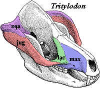
Eventually, of course, all of this sudden Permo-Triassic creativity collapses
into the mundane familiarity of the mammalian zygomatic arch, as in Tritylodon
or Probainognathus.
At this level, the jugal again is forced to give up its irresponsible behavior
and is harnessed to the exacting task of providing an attachment for the
powerful new masseter musculature. Rubidge
& Sidor (2001).
Therapsida:
Certainly, by the level of the therapsids, the jugal-squamosal connection is
well-established. See the images at the Profusely
Illustrated Guide. In fact, the squamosal seems to have developed by a
sort of competitive exclusion of the quadratojugal, which eventually disappears
entirely, with all of its functions being gradually subsumed by the
squamosal. The interesting part here is that we may speculate that the
jugal seems attracted (in some unspecified sense) to the squamosal generally,
not to some particular part or functionality. In this connection, note that the squamosal is a large,
plate-like bone in synapsids which
seems to have rather vague parameters. Unlike the jugal, it doesn't just
connect things. It covers area. If these generalizations are
meaningful we may suspect that the establishment of the jugal's squamosal
connection, together with the elimination of the constraining connection with
the quadratojugal, created a genetic condition in which the jugal had a lot of
freedom. That is, in any case, what we actually observe in the therapsid
lineage. The weird bulbous projections of Estemmenosuchus,
the quasi- vertical jugal of Tapinocaninus,
and the almost incomprehensible Lystrosaurus.
Eventually, of course, all of this sudden Permo-Triassic creativity collapses
into the mundane familiarity of the mammalian zygomatic arch, as in Tritylodon
or Probainognathus.
At this level, the jugal again is forced to give up its irresponsible behavior
and is harnessed to the exacting task of providing an attachment for the
powerful new masseter musculature. Rubidge
& Sidor (2001).
Mammaliformes: But, if that is the case, how do we derive the jugal of the Allotheria or, if one happens to be an unbeliever in this clade, the Multituberculata? Here, the jugal is reduced almost to a sliver wrapped up on the median side of a zygomatic process of the maxilla, very much like one of the postdentary bones being swallowed up by the dentary in contemporary cynodonts. Contrast this condition with the basal mammaliform Morganucodon, in which the zygomatic arch is dominated by the jugal. Kermack et al. (1981). The logical answer is that such a transformation, from the starting point of Tritylodon, is not very likely. In that event, the Allotheria (or Multituberculata, as the case may be) are more rationally considered a separate derivation from within the Cynodontia. Thus, the Mammaliformes, as commonly understood, may be polyphyletic.
Mammalia: However, we could still be well offside in jumping to this conclusion. The jugal seems to retain a good deal of plasticity in later forms. Thus, for example, the jugal is reduced or absent in the Monotremata and Insectivora, while, in the Didelphimorphia and Hyracoidea, it becomes so long that it actually contributes to the jaw articulation. It is likely that there is a strong correlation here with the development of the masseters and lateral jaw movements. So, for example, the jugals are particularly stout and well developed in the Rodentia, while strongly reduced in groups that do not chew or gnaw in the way of rodents.
The jugal is an interesting exercise because our information is good enough that we can attack issues such as: what has really made a difference in the evolution of this bone? We can make a short list as follows:
1. transformation from a sclerotic ring element to a circumorbital bone in contact with other dermal bones;
2. stabilization on the skull, probably by a relationship to the maxilla (recall that the reorganization of the maxilla in actinopterygians is associated with destabilization of the jugal);
3. loss of contact with the preopercular and contact with the quadratojugal and squamosal. This (a) may have been associated with a duplication of the jugal and (b) seems to have introduced some degree of instability in the posterior connections of the jugal, as seen in the anapsids;
4. stabilization of the jugal on the ventral margin of the skull in the reptilomorphs, which, oddly enough, seems to be associated with fixing its position relative to the orbit (compare the condition in the temnospondyls);
5. fenestration of the skull, resolving the posterior connection in favor of the quadratojugal, in diapsids, or the squamosal, in synapsids;
6. a number of, often homoplastic, changes in form related to vegetarianism without much real change in the underlying osteological relationships;
7. gradual specialization and fixation of the jugal as an important element of the zygomatic arch (note that this results in loss of the palatal contact);
8. a growing functional interdependence on the masseter musculature, with reduction or loss of the jugal associated with loss or reduction of this musculature.
It is hard to derive too much guidance from these generalizations. However, it does seem, once again, that the classical anatomists were correct. As in the business world, one's contacts make all the difference. It takes a very significant functional reorganization to disturb the relative stability of the fundamental osteological relationships. And, as we first observed in the premaxilla, in increased number of separate contacts results in progressive stabilization.
ATW020803. Last modified ATW040809
| Page Back | Unit Home | Glossary | Page Top | Page Next |
checked ATW040824