| Palaeos: |  |
Bones: The Gill Arches |
| Vertebrates | Overview |
| Page Back | Unit Back | Unit Home | Unit References | Unit Dendrograms ("Cladograms") |
Glossary | Taxon Index | Time |
| Page Next | Unit Next | Vertebrates Home | Vertebrate References | Vertebrate Dendrograms | Bones | Essay Index | Geography |
|
Bones
|
Gill Arches
Overview
|
 We will begin and, until later revision, also end
with a brief discussion of gill arches. The working hypothesis
here will be that the primitive jaw, consisting of the
palatoquadrate and Meckelian cartilage,
is derived from a hypothetical ancestral "mandibular
arch" and that the hyomandibular and related elements
are derived from a second (and much less hypothetical) hyoid
arch. Elements derived from the hyoid arch are most
conspicuously present in jaw support. Opinions vary on the
mandibular arch, and it may still be that the jaw has some
completely different ancestral homologue. However, the gill arch
theory is not only elegant, but has the virtue of being easy to
learn and remember, which may be why it has dominated the
discussion for the last century or so.
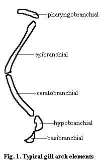
The segments of a typical gnathostome gill arch are shown in Figure 1. By convention, the corresponding parts of the hyoid arch are named by using the suffix -hyal, as in ceratohyal or basihyal, with the exception of the epihyal, which is more commonly called the hyomandibular, columella or stapes, depending on the subject matter and taxon under consideration and how badly the writer wishes to confuse you. Only the ceratal and epal elements of the mandibular arch are known. By other perverse conventions, the bone which would be the "epimandibular" is referred to as the palatoquadrate and the presumed "ceratomandibular" is Meckel's cartilage. The latter is named after Johann Friedrich Meckel (1781 - 1833), a Prussian physician and anatomist who had an abnormal fascination with abnormal human physiology*.
The phylogeny of the gill arches is of intense interest because of their possible involvement in the development of both the jaws and the paired appendages. For the past few years, the near-consensus has been that basal craniates had gills that were supported by a branchial basket, if they were supported at all. The branchial basket was braced against the body wall and unjointed, as in lampreys. It is derived from the hypomeres ( = lateral plate mesoderm). By contrast, the gill arches of gnathostomes are internal, jointed, and derived from the epimeres via the mesenchyme (i.e. with an admixture of neural crest ectoderm). This embryonic origin of the gill arches has been thought to be a good argument for their involvement with the jaw, but against their relationship with the limbs.
The recent discovery of the Cambrian craniate Haikouichthys (Chen et al. (1999)) has confused matters somewhat. Whatever else Haikouichthys may be, it is certainly not a gnathostome. It appears (study Chen et al.'s Figure 4 carefully) to have external gill arches (7?), as expected. However, (a) the gill arches seem to be jointed and (b) they appear to be closely related to paired fin-folds on the anaspid model. Indeed, Chen's cladogram places Haikouichthys basal to the anaspids and very close to the lampreys and Jamoytius. Thus, the possibility exists that the paired limbs of gnathostomes are derived from the ancient, external gill arches which have otherwise completely disappeared outside of the lampreys.
ATW 001118.
* This is, of course, is a very unfair summary of the life of an interesting scholar who studied with Cuvier and seems to have done a great deal of useful work in a variety of areas. Unlike many anatomists of the age, he made a study of vertebrate soft tissues and (according to one off-hand reference which gave no details) actually had the temerity to question Special Creation.
| Page Back | Unit Home | Glossary | Page Top | Page Next |
checked ATW030122