| Palaeos: | Bones: The Braincase | |
| Vertebrates | The Pleurosphenoid |
| Page Back | Unit Back | Unit Home | Unit References | Unit Dendrograms ("Cladograms") |
Glossary | Taxon Index |
| Page Next | Unit Next | Vertebrates Home | Vertebrate References | Vertebrate Dendrograms | Bones | Time |
|
Bones
|
Braincase
Overview |
Sphenoid Region
Overview |

About three years ago (1999) we posted a small paragraph on the pleurosphenoid at the bottom of the Braincase Overview page. Here, we essentially accused the profession of having made the whole thing up. We received several emails from pleurosphenoid afficionados objecting that one or the other reptile actually had this no-doubt essential piece of vertebrate headgear. Eventually (2001), we grudgingly admitted that at least some Gymnophiona had a pleurosphenoid. See the Grandisonia braincase at right. More recently, we dealt with the evolution of the jaw in Holocephali, as illuminated by Grogan et al. (1999). This important article leads us back to the middle of the braincase and a re-evaluation of a number of structures, including the pleurosphenoid. While we are not yet willing to yield the pleurosphenoid a seat at the High Table of serious cranial structures, we can at least relieve it of the singular indignity of being a footnote among head bones.
 Overview
Unlike many cranial bones, the function of the pleurosphenoid is easier to determine than its structure or lineage. In gnathostomes generally the anterior braincase is the province of a sphenethmoid ossification which develops, more or less, from the trabeculae. However, something rather dramatic occurred in the tetrapod lineage during the later part of the Early Carboniferous. At this point, temnospondyls, lepospondyls and reptilomorphs diverged. None of these taxa has a "normal" gnathostome sphenethmoid, although the lepospondyls come close. In most cases, the anterior braincase is unossified, or the degree of ossification is reduced, presumably as a result of adapting to a terrestrial or partially terrestrial existence.
This leaves things like crocodiles, theropods, and even birds and mammals, with a real problem. The braincase can be made cartilagenous without a great deal lost. However, it is still necessary to shore up the sides of the skull if one is to develop a really formidable rostrum. Furthermore, it is important to keep things like eyeballs, maxillary veins, and nasal epithelia from sloshing around and generally getting in each others' business. Thus, many tetrapod taxa re-invented the sidewall for the anterior braincase, or at least bits and pieces of it.
The pleurosphenoid is oneof those bits and pieces. It is distinguished (if at all) by its position relative to certain embryological markers, notably the pila antotica. On the off-chance that some particularly obtuse or forgetful reader may be unfamiliar with this ever-popular landmark of lizard embryology, we will explain.
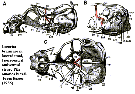 The sphenethmoid does not simply disappear over the course of tetrapod
evolution. Rather, like a decrepit dwelling, it gradually sheds
superstructure until only a few main beams remain. In development, the
embryo still dutifully constructs these beams as a latticework of cartilage,
although the frame will never be built out or ossified. One of these beams
is the pila antotica. These pilae [1] (since there
are actually a pair) arise from either side of the dorsum
sellae, the sturdy process which guards the pituitary fossa just in
front of the hind-brain. With reference to our standard diagram, the dorsum
sellae is located just about at the point where the polar cartilages are
shown. Dorsoventrally, it is more or less at the bottom of the
brain. The pilae rise anteriorly and medially from the dorsum sellae
until the two processes meet in the middle. It then rises to the taenia
marginalis which runs sagitally along the midline of the skull table, just
under the dermal bones.
The sphenethmoid does not simply disappear over the course of tetrapod
evolution. Rather, like a decrepit dwelling, it gradually sheds
superstructure until only a few main beams remain. In development, the
embryo still dutifully constructs these beams as a latticework of cartilage,
although the frame will never be built out or ossified. One of these beams
is the pila antotica. These pilae [1] (since there
are actually a pair) arise from either side of the dorsum
sellae, the sturdy process which guards the pituitary fossa just in
front of the hind-brain. With reference to our standard diagram, the dorsum
sellae is located just about at the point where the polar cartilages are
shown. Dorsoventrally, it is more or less at the bottom of the
brain. The pilae rise anteriorly and medially from the dorsum sellae
until the two processes meet in the middle. It then rises to the taenia
marginalis which runs sagitally along the midline of the skull table, just
under the dermal bones.
In some forms, as in Multituberculata, the pila antotica itself may ossify. In such cases, it is referred to as the taenia clino-orbitalis [2]. More frequently, as in birds, crocs, and ocasional others, the pilae allegedly come to support small and variable ossification above, more or less as minor anterior extensions of the otic capsule. These, we are told, are the pleurosphenoids.
And so, although we are still inclined to suspect that we have been taken in by some bizarre anatomical jest, we have done full justice to the subject and will say no more. ATW020917.
[1] Pila is normally translated as pillar, but is more acurately a generic Latin word for any long, vaguely cylindrical object. It also has a few other meanings, including ball, ball game, and bookstore. Thus: pilam pilarum inter pilas pilae ludabamus (we were playing a game with balls among the pillars of the bookstore).
[2] No, I am not kidding.
| Page Back | Unit Home | Glossary | Page Top | Page Next |
checked ATW060127