| Palaeos: | Bones: The Gill Arches | |
| Vertebrates | The Hypohyal |
| Page Back | Unit Back | Unit Home | Unit References | Unit Dendrograms ("Cladograms") |
Glossary | Taxon Index | Time |
| Page Next | Unit Next | Vertebrates Home | Vertebrate References | Vertebrate Dendrograms | Bones | Essay Index | Geography |
|
Bones
|
Gill Arches
Overview
|
 The hypohyal is a ventral element of the hyoid arch which links the ceratohyal
and the basihyal. If your reaction to this, "well, of course!"
then you probably don't need to be reading this page. For those who did not
imbibe splanchnocranial anatomy with their mother's milk, we will return to
fundamentals.
The hypohyal is a ventral element of the hyoid arch which links the ceratohyal
and the basihyal. If your reaction to this, "well, of course!"
then you probably don't need to be reading this page. For those who did not
imbibe splanchnocranial anatomy with their mother's milk, we will return to
fundamentals.
The gill arches have two basic parts, dorsal and ventral. Each part is associated with a main gill arch segment, the (dorsal) epal and (ventral) ceratal segments, respectively. Thus, each of the branchial arches, the arches which actually function as respiratory arches in fishes, has an epibranchial and a ceratobranchial. The hyoid arch is a an additional arch anterior to the first branchial arch. The hyoid is often involved in respiration, but its primary functions are related to jaw support and extension. The ceratal and epal segments of this arch are called the ceratohyal and the hyomandibula, respectively.
Most gnathostomes have some kind of ventral extension of the gill arches, a basal component, and these usually lie flat along the ventral "throat" of the fish. The basal elements of adjacent arches are often closely interlocked, or even fused. The basal element of the hyoid arch, the basihyal, is no exception.
 Now,
the ceratal segment is typically long and relatively rigid and at least slightly
vertical, while the basal elements are typically fused and/or tightly bound to
the base of the gullet in a horizontal orientation. Keeping these elements
in articulation as they move in different planes requires a complex joint.
This is normally supplied by a small element of complex shape. This
element is the hypobranchial
or, in the case of the hyoid arch, the hypohyal.
Now,
the ceratal segment is typically long and relatively rigid and at least slightly
vertical, while the basal elements are typically fused and/or tightly bound to
the base of the gullet in a horizontal orientation. Keeping these elements
in articulation as they move in different planes requires a complex joint.
This is normally supplied by a small element of complex shape. This
element is the hypobranchial
or, in the case of the hyoid arch, the hypohyal.
We may define the Standard Condition by reference to the amiable Amia calva. I say "by reference to" since Amia itself has some relatively non-standard features. However, the excellent images of Grande & Bemis (1998) dictate our choice. Finding good images of the hypohyal is, as we have discovered, not a simple proposition.
Note that the hypohyal in Amia seems to have a rather solid and immobile articulation with the ceratohyal. In fact, the articular surface is cartilagenous. However, most of the mobility in the hypohyal is in the contact between the hypohyal and the basibranchials. Amia departs from the Standard Condition, as we will define it, in that there is no separate basihyal. The hypohyals directly contact the basibranchials, which are fused into a more or less continuous "basibranchial copula," although the basibranchials are only partially ossified. The hypohyals are also seldom completely ossified. Even in the rather large individual shown in the image, the hypohyal remains partially cartilagenous. This is a typical condition in actinopterygians.
 The position of the hypohyals shown in the image is not life-like. In
life, the first 3-5 branchiostegal rays of each side overlap their counterparts
on the opposite side. This is possible because the ceratohyals are
strongly curved as shown in the occipital view in the figure at right. Accordingly, the
hypohyals must be directed amost ventrally. In this orientation, the articular "head" of the
hypohyal probably articulates with the hypobranchials of Branchial Arch I as
well as with the basibranchial copula. It also seems likely that the two
hypohyals also make rolling contact with each other in the process of raising and
lowering the floor of the pharyngeal cavity via the branchiostegal
rays.
The position of the hypohyals shown in the image is not life-like. In
life, the first 3-5 branchiostegal rays of each side overlap their counterparts
on the opposite side. This is possible because the ceratohyals are
strongly curved as shown in the occipital view in the figure at right. Accordingly, the
hypohyals must be directed amost ventrally. In this orientation, the articular "head" of the
hypohyal probably articulates with the hypobranchials of Branchial Arch I as
well as with the basibranchial copula. It also seems likely that the two
hypohyals also make rolling contact with each other in the process of raising and
lowering the floor of the pharyngeal cavity via the branchiostegal
rays.
Janvier (1996) takes the hypohyal to be general for the Gnathostomata. This is unclear. The ventral branchial arch segments of placoderms are so poorly known that nothing useful can be said. Apparantly, placoderm ventral arch elements were almost entirely cartilagenous. In at least many Chondrichthyes, the hypobranchials are well developed, although they point posteriorly. However, there is no hypohyal. The ceratohyal articulates directly with the basihyal or basibranchial copula. Thus, we might suppose that the hypohyal is a special feature connected with the development of the branchiostegal apparatus.
 This
suspicion is confirmed from at least some reconstructions of the hyoid arch in Acanthodii.
The branchial arches in the Acanthodii are not well ossified, so the issue is
murky. The ceratobranchials tend to have two ossification centers.
What seems to be unsettled is whether this is also true of the ceratohyal, so
that the more ventral of the two ceratohyal segments becomes the hypohyal, or
whether both segments are parts of the ceratohyal and the hypohyal was inserted
later, as a neomorph. In the latter case, the hypohyal is possibly
polyphyletic, i.e. convergently developed in actinopterygians and
sarcopterygians.
This
suspicion is confirmed from at least some reconstructions of the hyoid arch in Acanthodii.
The branchial arches in the Acanthodii are not well ossified, so the issue is
murky. The ceratobranchials tend to have two ossification centers.
What seems to be unsettled is whether this is also true of the ceratohyal, so
that the more ventral of the two ceratohyal segments becomes the hypohyal, or
whether both segments are parts of the ceratohyal and the hypohyal was inserted
later, as a neomorph. In the latter case, the hypohyal is possibly
polyphyletic, i.e. convergently developed in actinopterygians and
sarcopterygians.
Neither one of these possibilities is particularly appealing. But, of the two, we tend to prefer the former on morphological grounds. The Late Devonian actinopterygian Mimia, for example, has a well-preserved hypohyal which looks quite like the ceratohyal, as in Acanthodes. It is only as we approach the Neopterygii and the Standard Condition that the hypohyal becomes short, rounded and joint-like. Development beyond the basal Neopterygii is a matter of adding an additional hypohyal -- presumably increasing speed, flexibility and control of the movement of the branchiostegal apparatus as teleosts developed more and more elaborate specializations related to suction feeding. See, e.g., image at Novumbra.
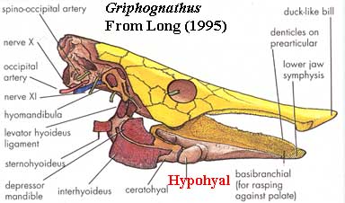 On
the other side of the teleostome divide lie the the sarcopterygians. Here,
and particularly in the durophagous forms, the ceratohyal and an anterior
extension of the basibranchial copula become strongly involved in supporting the
lower jaw, rather than in lowering the floor of the mouth. So, in Glyptolepis,
for example, the ceratohyals are rather large, flat elements supporting the
lower jaw and the hypohyals are correspondingly stout joints providing the
necessary flexibility between the ceratohyals and the fused
basibranchials. This trend reaches something of a logical extreme in Dipnoi,
such as Griphognathus, in which the basibranchial copula is the
lower jaw and the hypohyal effectively takes the place of the articular. Long
(1995).
On
the other side of the teleostome divide lie the the sarcopterygians. Here,
and particularly in the durophagous forms, the ceratohyal and an anterior
extension of the basibranchial copula become strongly involved in supporting the
lower jaw, rather than in lowering the floor of the mouth. So, in Glyptolepis,
for example, the ceratohyals are rather large, flat elements supporting the
lower jaw and the hypohyals are correspondingly stout joints providing the
necessary flexibility between the ceratohyals and the fused
basibranchials. This trend reaches something of a logical extreme in Dipnoi,
such as Griphognathus, in which the basibranchial copula is the
lower jaw and the hypohyal effectively takes the place of the articular. Long
(1995).
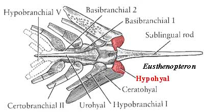 Our
own inheritence from the basal sarcopterygian tradition is a bit less extreme,
as can be seen in the branchial apparatus of Eusthenopteron, shown at
left in a figure from Carroll (1988).
The basibranchial copula has been drawn anteriorly with a long, but slim,
sublingual rod. The hypohyal is round and stout, providing an articular
bearing between both the sublingual rod and the copula proper, on the one hand,
and the ceratohyal, on the other. Note that, contrary to the Standard
Condition, the hypohyal appears to have a fixed contact on the copula and a
moving contact with the ceratohyal.
Our
own inheritence from the basal sarcopterygian tradition is a bit less extreme,
as can be seen in the branchial apparatus of Eusthenopteron, shown at
left in a figure from Carroll (1988).
The basibranchial copula has been drawn anteriorly with a long, but slim,
sublingual rod. The hypohyal is round and stout, providing an articular
bearing between both the sublingual rod and the copula proper, on the one hand,
and the ceratohyal, on the other. Note that, contrary to the Standard
Condition, the hypohyal appears to have a fixed contact on the copula and a
moving contact with the ceratohyal.
This conformation remains oddly constant over the loss of functional gills in the Tetrapods. Unfortunately the hyoid apparatus is almost invariably unossified and, worse, the terminology changes. The furthest we have been able to go with a hypohyal, eo nomine, is the salamander Necturus. The hyoid apparatus of Necturus looks quite a bit like a truncated version of Eusthenopteron, without the sublingual rod and with only the hyoid and first branchial arches. Since the sublingual rod is missing, the two hypohyals meet medially and cap the the basibranchial copula.
 Amniotes
also have a hyoid apparatus. Some examples are shown in the figure from Romer
(1956). It appears, in the case of Heloderma that
there is a separate cartilage between the ceratohyal and the basibranchial
body. This may or may not be homologus to the hypohyal. However, in
almost all other taxa, the ceratohyal merges indistinguishably with the
copula. In the synapsid lineage, the pieces are perhaps better
differentiated, but mammallian anatomy has developed its nomenclature along a
very different path, and the homologies are, in any case, far from clear.
ATW030328
Amniotes
also have a hyoid apparatus. Some examples are shown in the figure from Romer
(1956). It appears, in the case of Heloderma that
there is a separate cartilage between the ceratohyal and the basibranchial
body. This may or may not be homologus to the hypohyal. However, in
almost all other taxa, the ceratohyal merges indistinguishably with the
copula. In the synapsid lineage, the pieces are perhaps better
differentiated, but mammallian anatomy has developed its nomenclature along a
very different path, and the homologies are, in any case, far from clear.
ATW030328
| Page Back | Unit Home | Glossary | Page Top | Page Next |
checked ATW030122