| Palaeos: |  |
Bones: The Ear |
| Vertebrates | The Incus |
| Page Back | Unit Back | Unit Home | Unit References | Unit Dendrograms ("Cladograms") |
Glossary | Taxon Index |
| Page Next | Unit Next | Vertebrates Home | Vertebrate References | Vertebrate Dendrograms | Bones | Time |
| Bones Braincase Dermal Bones Ear Gill Arches Teeth |
Ear Overview The Incus References |
The incus is the first of the palatoquadrate derivatives we will take up. As we will discuss if, as, and when the palatoquadrate essay is ever written, the hypothetical first gill arch of chordates (or, alternatively, the velar skeleton) begat the palatoquadrate and Meckel's cartilage. The palatoquadrate begat a number of things, but most importantly the epipterygoid and the quadrate. The quadrate, in turn, begat the incus.
Thus, the ossification which became the incus has served, in one taxon or another, as a support element in filter feeding and possibly respiration, as a structural part of the occiput, the cheek, and the braincase, and as a critical mechanical element in the jaw and in hearing. Despite this enormous variety of functions, in thousands of vertebrate families and over five hundred million years of evolution, this bone has retained an articulation with Meckel's cartilage, or its progeny, the articular and the maleus. Over all or most of that same range (depending on your understanding of the origin of jaws), it has almost always retained a separate articulation with the hyomandibula or its progeny, the stapes and columella. If we are ever to develop a theory of vertebrate osteology, this is one of the most remarkable facts it will have to explain.
 Fortunately,
our task here is much simpler: to give a brief account of the middle bone of the
three mammalian auditory ossicles. In reptiles with auditory ossicles, the
entire job is done by the stapes or columella, with or without an accessory
extracolumella. Only in mammals are there three ossicles: the maleus, incus, and
stapes. There is undoubtedly a great and interesting body of literature on the
functional anatomy of the ossicles, but we will have to be content with bare
structure for the moment.
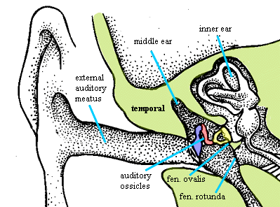
As an overview and reminder, at right is schematic vertical section of the human ear adapted from that most excellent of overviewers and reminderers, Kardong (1998: 659). Some additional background information (and probably misinformation) can be gleaned from The Ear. Briefly, Sound vibrations in air are funnelled down the external auditory meatus, at the end of which is the tympanic membrane. Having no place else to go, the pressure waves in the air cause the tympanic membrane to flutter. The movement of the membrane is picked up by the maleus (blue), which is in direct contact with the tympanum, and pased through the incus (red), and finally the stapes (yellow). The stapes has a large footplate which fits into the oval window of the inner ear and transduces the physical movement of the bones back into pressure waves in the perilymph of the inner ear. These pressure waves are again transduced into electrical impulses by cells in the inner ear complex related to the ancient lateral line system of fishes. Ultimately (i.e. microseconds later), we perceive the electrical disturbance as sound.
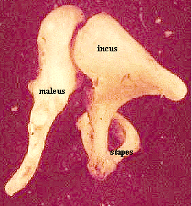
 A
more realistic view of the ossicles in articulation is shown at left. It is
actually the long process of the maleus, the manubrium, whch makes the
primary contact with the tympanum. The manubrium is, in fact, the remote
ancestor of the retroarticular process of the articular. The maleus makes
contact with a broad facet on the head of the incus. The incus has a lenticular
process or crus longum which articulates with the stapes.
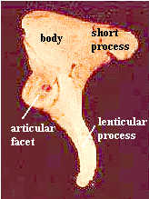
 The geography of the incus is shown in a bit more detail at right. The articular
facet (the incudomalleal joint) is close to the lenticular process. The joint faces anteromedially (except in whales) and is freely moveable on a cartilagenous surface -- not a sutural connection.
The short process, or crus breve, is the attachment point for a ligament which binds it to the
wall of the epitympanic recess, that is, the end of the middle ear opposite the
Eustachian tube. Another ligament attaches to the body of the incus and binds it
to the roof of the middle ear (tegmen tympani). Thus the movement of
the incus inresponse to the maleus is probably constrained fairly tightly,
although one can't tell just from the anatomy whether it is effectively
constrianed to one dimension. Perhaps, like the loose quadrate-articular joint
from which it evolved, there is significant play in at least two planes.
The joint between the incus and stapes is likewise a cartilagenous joint, with a tendency to ossify in older humans. Again, the implication is that the movement at this joint is constrained, but not necessarily confined to stereotyped motion in single plane. In fact, evolution seems to have avoided this solution -- one it could easily have been acheived, given the quadrate as starting material. This may have important implications for the hearing. A question raised before in these Notes, and not yet answered, is whether this system is delivering one, or two-dimensional information to the inner ear. It is not necessary that the tympanum react as a one-dimensional ocillator or, even if it does, that it vibrate only to a single frequency to the exclusion of, for example, harmonics. Obviously mammals are capable of hearing more than one frequency at a time. This could be accomplished by decomposing a complex one-dimensional wave representing the superposition of all frequencies. However, it is equally possible that some of the decomposition is performed mechanically, before the impulse is transduced to the inner ear. Recall that the lateral line mechanoreceptors are exquisitely sensitive to the direction, as well as the magnitude, of shearing forces. The more elegant solution might be to evolve a system which takes advantage of this feature by supplying a vector signal to the inner ear: one with direction as well as magnitude. However, this is all speculation, and we must leave the matter to the physiologists with real data.
One peculiarity of the incus is that, at least according to some sources, not all of the bone is derived from the quadrate, or from any part of the first pharyngeal (mandibular) arch. The lenticular process is actually derived from mesenchyme attributed to the second branchial (hyoid). perhaps it may more appropriately be viewed as a part of the stapes, rather than the incus. Thus, from an evolutionary perspective, the lenticular process may be homologous to the shaft of the stapes and result from fusion of the former quadrate- stapes articulation. This speculation is consistent with the observation that artiodactyls have a very short lenticular process (Thewissen & Hussain (1993)), with perhaps greater length to the corresponding process of the stapes.
The most obvious matter has been left for last, i.e. why bother with this complex arrangement? At least with regard to the incus, the answer seems to relate to amplification. The lenticular process is substantially longer than the arm of the manubrium which connects the tympanum to the articular facet. The result is that displacement of the malleus is amplified because it results in a larger displacement of the lenticular process. In essence, the incus acts as a lever with the body as the fulcrum, held steady by the ligamentous connections described above. This explains why the body, whichis not involved in sound conduction, is so much more massive and compact than the lenticular process. Its function is precisely not to move, but to hold the system steady. See Ear Physiology* for a rather 19th century-style diagram of the system. This is only one of several amplification and control mechanisms in the middle ear. However, the others relate to the malleus and stapes. ATW030125.
Howstuffworks \How
Hearing Works\ basic mechanics, with some oversimlification of certain
points.
Bio203 the ear: pictures of large plaster models.
PICTURES OF OSSICULAR CHAIN RECONSTRUCTION, INCUS REPLACEMENT, ...
better, but mostly concerned with prosthetics
| Page Back | Unit Home | Glossary | Page Top | Page Next |
checked ATW050819