| Palaeos |  |
Archosauria |
| Vertebrates | 670: Prestosuchidae (2) |
| Page Back | Unit Home | Unit Dendrogram | Unit References | Taxon Index | Page Next |
| Unit Back | Vertebrates Home | Vertebrate Dendrograms | Vertebrate References | Glossary | Unit Next |
|
Abbreviated Dendrogram
ARCHOSAUROMORPHA | `--ARCHOSAURIA |--Proterosuchidae `--+--Erythrosuchidae `--+--Euparkeriidae `--Crown Group Archosauria |--Ornithodira | |--PTEROSAURIA | `--DINOSAUROMORPHA `--Crurotarsi |--Phytosauridae `--Rauisuchiformes |--Rauisuchia | |--Ornithosuchidae | `--+--Prestosuchidae | | |--Saurosuchus | | `--+--Ticinosuchus | | `--Batrachotomus | `--Rauisuchidae `--Suchia |--Aetosauridae `--CROCODYLOMORPHA |
Contents
Overview |
 |
|
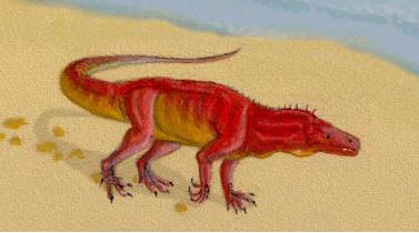
Ticinosuchus ferox as it may have appeared in life, length about 2.5 meters. Anisian/Ladinian of the north Tethys shoreline. These animals, collectively called Rauisuchia or Prestosuchidae, seem to represent the central line of archosaur evolution during the Middle Triassic. They were active, terrestrial, carnivorous animals with an efficient fully erect gait; not unlike a sort of terrestrial crocodile which erect rather than sprawling (semi-erect) stance. Whilst some like Ticinosuchus were modest-sized animals, others attained as much as 6 meters or more in length. Because of their terrestrial lifestyle, and because big predators tend to be less common than herbivores and smaller animals, the "Prestosuchia" are poorly represented in the fossil record, and the precise phylogenetic relationships of the group remain disputed. Image in A. H. Müller, Lehrbuch der Paläozoologie, p.296, after Krebs 1965. |
 Ticinosuchus: T. ferox Krebs, 1965; "Mandasuchus"
Range: Middle Triassic (Anisian and Ladinian) of Europe and Africa
Phylogeny: Prestosuchidae :: Batrachotomus + *.
Comments: A partial skeleton of Ticinosuchus ferox is known from the Grenzbitumenzone (Anisian-Ladinian boundary), Tessin River, near Monte San Giorgio, Canton Tessin, Switzerland. However, this animal is also known from the Ladinian of northern Italy. At 2.5 metres it filled the medium terrestrial predator niche. Fossilized footprints called Chirotherium "hand beast" have also been found. They were probably left by an animal very like this one.
Characters: vertebral intercentra absent [P93]; cervical vertebrae elongate [S74]; calcaneal tuber with lateral side concave & medial side convex [P93]; rotary, crurotarsal ankle joint present [P93]; one pair of paramedian armor plates per vertebral segment [$P93].
Image: Ticinosuchus, courtesy (not to mention considerable tact and patience) of Daniel Bensen.
Links: Ticinosuchus (German)
References: Parrish (1993) [P93], Sill (1974) [S74]. MAK991019 and 030808, ATW031129.
 Batrachotomus:
Batrachotomus kupferzellensis Gower
Range: Late Ladinian (Lettenkeuper) of Germany
Phylogeny: Prestosuchidae :: Ticinosuchus + *.
Comments: The dominant predator of the Lower Keuper environment, Batrachotomus was an enormous animal, up to 6 meters long (the size of Saurosuchus) It is well-known by teeth, partial skeletons and cranial material, and resembled other large archosaurs like Ticinosuchus, Saurosuchus, and Postosuchus. Cladistic analysis by Benton & Walker (2002) gives a very close relationship between Batrachotomus and Prestosuchus, with Saurosuchus as the sister taxon of these two. This species may therefore be included within a monophyletic Prestosuchidae, or as a very close sister group.
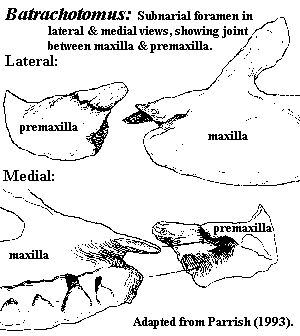
Characters: subnarial foramen with joint between maxilla &
premaxilla [P93] [1]; jugal excluded from antorbital fenestra [π$03];
basioccipital forming almost all of occipital
condyle [G02]; two pairs of basal tubera on basioccipital [G02]; basioccipital
almost or entirely excluded from foramen
magnum [G02]; supraoccipital triangular & much wider than long,
forming dorsal border of foramen magnum [G02]; supraoccipital, dorsal surface
with median ridge and depressed lateral facets for parietals [G02];
supraoccipital with posterolateral processes overlapping paroccipital processes
[G02]; exoccipitals & opisthotics fully fused [G02]; exoccipital with large
vertical crista
forming posterior margin of metotic
foramen [G02]; hypoglossal
foramen anterior to crista [G02] [2]; paroccipital processes
short & strongly expanded distally [G02]; metotic foramen not subdivided [3]
[G02]; epiotic
present as thin, irregular strip adjacent to supraoccipital facets for parietals
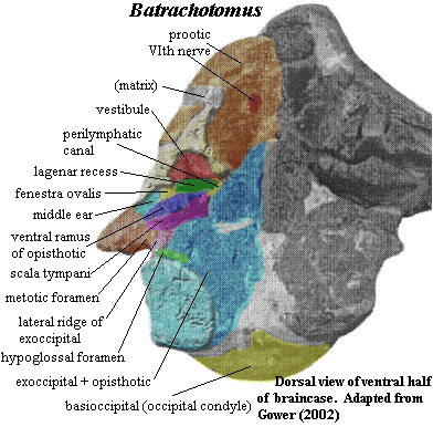
[G02]; inner ear with moderately well-developed lagenar
recess and ventral opisthotic ramus  separating scalae
vestibuli & tympani (see image [4]) [G02]; lagena not
partitioned from remainder of scala vestibuli (in contrast to crocodylomorphs)
[G02]; additional foramen (posterior cephalic vein?) passing into dorsal end of
metotic foramen [π$03]; epiotic, prootic & opisthotic meet in Y-shaped suture medial to
vestibule [G02]; perilymphatic foramen (into rec. scala tympani) small
compared to crocs & aetosaurs [G02]; crista
prootica slight [G02]; basisphenoid bends ventrally anterior to otic
capsule [G02] [5]; deep, blind, undivided basioccipital recess on ventral
surface between tubera, possibly homologous to crocodylian median
eustachian foramen (which communicates with pharynx) [G02]; basisphenoid
strongly waisted between tubera and basipterygoid
processes [G02]; entrance of internal
carotid artery on lateral face of basisphenoid in notch between basal
tubera of basisphenoid & basipterygoid process [G02]; Vidian
canal absent [G02]; external foramen for exit of abduscens nerve on
underside of horizontal surface [π$03] [6]; trigeminal
foramen at least partially divided by prootic from foramen for middle
cerebral vein [π$03]; laterosphenoid probably long and thin [G02]; vertebral intercentra absent [P93]; rotary, crurotarsal
ankle joint present [P93].
Notes: [1] Note the surprising reappearance of this character, which one associates with more basal taxa, such as the Proterosuchidae or perhaps the Ornithosuchidae. [2] The point is important, since it differentiates Batrachotomus from the Suchia. [3] For the significance of this, see discussion at the Inner Ear. The subdivision in question splits the metotic foramen into a vagus foramen (nerve exits) and a separate fenestra pseudorotunda of the recessus scala tympani. [4] it may help to copy the image and magnify it about 4X. [5] This effect has been exaggerated by preservational artifact, and its hard to tell how seriously we may take Gower's [G02] description of the fossil (entirely accurate as far as we know) as a description of the living animal's skeleton. He does not provide a reconstruction. [6] "[π03]" refers to our own analysis of published data.
Reference: Gower 2002 [G02], Parrish (1993) [P93]. ATW031122.
| Page Back | Unit Home | Page Top | Page Next |
checked ATW060119
Using this material. All material by ATW is public domain and may be freely used in any way (also any material jointly written by ATW and MAK). All material by MAK is licensed Creative Commons Attribution License Version 3.0, and may be freely used provided acknowedgement is given. All Wikipedia material is either Gnu Open Source or Creative Commons (see original Wikipedia page for details). Other graphics are copyright their respective owners