| Palaeos: |  |
Glossary |
| The Vertebrates | Eo-Ez |
| Page Back | Unit Back | Unit Home | Unit Dendrogram | Unit References | Glossary | Taxon Index |
| Page Next | Unit Next | Vertebrates Home | Vertebrate Dendrograms | Vertebrate References | Bones | Time |
For most phrases beginning with directional words, e.g. "posterior," "dorsal," "external," etc., or some generic anatomical terms, e.g., "vena," look under the next word in the phrase. However, note that this convention is not used with complete consistency in this Glossary.
Eocene: The second Epoch of the Cenozoic, 54.8-33.7 Mya. The Early Eocene (34.8-49 Mya) is the Ypressian Age. The Middle Eocene (49.0-37.0 Mya) includes the Lutetian and Bartonian Ages. The Late Eocene (37.0-33.7 Mya) is the Priabonian Age.
Epal: in fishes, relating to the upper main gill arch segment (i.e., the epibranchials, hyomandibula or palatoquadrate). See Gill Arches.
Epaxial: the region dorsal to the lateral septum in fishes. Although tetrapods lack a lateral septum, this region remains developmentally distinct and gives rise to a special set of muscles, the epaxial musculature, which includes elements ultimately traceable to the dorsal fin musculature of fishes. The lateral septum in fishes extends laterally from the spinal cord. The spine tends to be somewhat centrally located, and the epaxial region is thus quite large. In tetrapods the epaxial region is more limited.
Epiblast: see gastrulation.
Epibranchial: the more dorsal of the two main gill arch elements. Serial homologues of the palatoquadrate and the hyomandibula. See the Epibranchials.
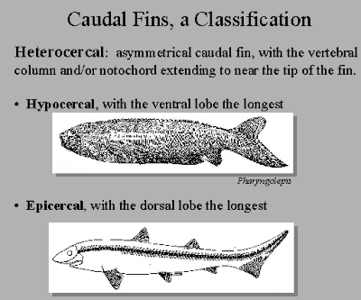
Epicaudal lobe: the part of the tail fin above the notochord.
 Epicercal: of the tail (caudal) fin of certain fishes,
upwardly tapering. Generally, this signifies that the notochord or spine forms
the upper margin of the tail fin. Same as heterocercal,
according to some. But see the image at right, which makes more sense to us.
Epihyal: this should be the same as the hyomandibular. That is, the hyomandibula is (very probably) the epal element of the hyoid arch. However, in many actinopterygian fishes, the ceratohyal (ceratal element of the hyoid arch) has two separate ossifications. The more proximal (or posterior or dorsal, depending on the fish and frame of reference) is frequently referred to as the epihyal.
Epidermis: the outermost layer of the integument (see figure at that entry) During development, the inner layer (stratum basale or stratum germinativum) is in contact with the basement membrane.
Epiglottis: A small thin flap of cartilage behind the tongue that covers the larynx during swallowing.
Epimere: See Early Development Terms.
Epineural: in fishes, a slender bone which lies in the myoseptum and projects backwards and upwards from the neural arch and spine. Epineurals may be forked. Compare supraneural.
Epiotic: a ?median dorsal ossification of the otic capsule, somewhat like a second, more anterior, supraoccipital. This bone, if present, normally has no external exposure. Presumably, like the supraoccipital, it arises by ossification of dorsal soft tissues joining the otic capsules dorsally.
Epiphysis: [1] the terminal, usually ossified sections of long bones, including the articulating surfaces. As opposed to the diaphysis and metaphyses. [2] a small, light-sensitive endocrine gland in the brain; situated beneath the back part of the corpus callosum which secretes elation.
Epipodial: same as epipodiale. See figure at metapodial
Epipodiale: (pl. epipodialia) one of the distal limb bones, i.e. ulna, radius tibia or fibula. See figure at metapodial.
 Epipterygoid:
The epipterygoid is a misnomer. Like the quadrates, the epipterygoids
are ossified portions of the palatoquadrate (the original upper jaw which, like
the hyomandibular, is homologous with an upper gill arch segment). The
epipterygoids often appear to arise from the pterygoid, but do not. The
epipterygoids are the true, old stuff of the palatoquadrate, while the pterygoid
is but common dermal bone with pretensions. In fact, the epipterygoids are
the original braincase articulations of the palatoquadrate. They
demonstrate this ancient nobility by rising up in a graceful curve to reach the
bones of the skull roof, like the last remaining columns of an abandoned
temple.
Epitegum: a shield element of jawless fishes, considered as arising from separate growth centers. The ventral shield is considered as a single epitegum.
Epithelium: a tissue forming the boundary of an organ; a characteristic tissue type forming such boundaries.
Epitympanic recess: the distal or upper end of the middle ear, opposite the Eustachian tube. The middle ear ossicles of mammals are lodged in this part of the middle ear.
Epoccipital: one of the small bones along the margin of the parietal frill of many Ceratopsia.
Epural: in fish tail anatomy, an elongate detached bone above the urostyle and behind the last neural spine supporting caudal fin rays. Apparently derived from neural spines or the urostylic centra; dorsal homologues of the hypurals. Vary in number between one in advanced fishes to three in primitive actinopterygians. From Dictionary of Ichthyology (site no longer available). See image at urodermal.
Eretmo-: Greek root for oar.
Erythro-: Greek root for red.
Escuminac Formation: Frasnian? (Late Devonian) of Canada. This is the formation containing the famous Miguasha fauna including many incomparable specimens of Eusthenopteron, possibly the best known Middle Paleozoic vertebrate. The sediments are probably coastal marine or possibly deltaic, although they are frequently reported as fresh water in the older literature. Schultze & Arsenault (1985).
Ethmoid: Gr. ethmos = a sieve, and eidos = resemblance. A term describing the structure of the ethmoid bone of the skull, the most anterior of the four principal braincase regions. It is associated with the nasal capsules and chemosensation. Frequently it fuses with the sphenoid region and is referred to as the sphenethmoid. In mammals, the ethmoid is reduced to a series of turbinals: very thin bones in the nasal passage that help recover respiratory water vapor.
Ethmoid articulation: of the palatoquadrate. An anterior articulation of the palatoquadrate in which the palatoquadrate articulates with a an anterior portion of the sphenethmoid portion of the braincase. See image at paratemporal articulation.
Ethmosphenoid: same as sphenethmoid. The combined sphenoid and ethmoid regions of the braincase. The anterior half of the braincase, physically separate from the posterior, otoccipital unit in Sarcopterygii. See Bones: The Braincase.
Euautostylic: jaw suspension in which the jaw is attached directly to the braincase as in placoderms.
Eurybasal: having a wide fin base (as sarcopterygian fin); opposite of stenobasal.
Euryhaline: tolerating a wide range of salinity; opposite of stenohaline.
Eustachian tube: a passage joining the middle ear and pharynx.
Eutaxy (= Eutaxis): of bird wings, having the 5th secondary present. Adj. Eutaxic. Opposite of diastaxy.
Euxinic: relating to a water layer or water column which is anoxic (little or no oxygen) and sulfidic (contains reduced sulfur species, e.g. H2S, FeS, etc., as opposed to sulfate). In Phanerozoic environments, euxinic waters are generally deep, stagnant, and cool, since any significant current or convection would tend to introduce oxygen. The best modern example is the bottom of the Black Sea. In fact, the term is derived from the Greek name for the Black Sea, Εὔξεινος Πόντος.
Evaporite: a deposit created by the evaporation of sea water.
Exaenodont: in mammalian dentition, a condition of the cheek teeth in which the base of the crown is significantly lower on the buccal than on the lingual side of the tooth. Rich et al. (2001).
Exapt: to adapt, by selection, to a different purpose. Examples: (1) the muscles homologous to the usual vertebrate eye muscles have become exapted to move the tentacles in caecilians; (2) the swim bladder, originally adapted for control of buoyancy, was exapted as a respiratory organ in various groups of fish.
Exoccipital: The exoccipitals are paired bones of the occiput. They derive from the neural arch elements of embryonic vertebrae which have been incorporated into the braincase. Dorsally, the exoccipitals contact the supraoccipital and the foramen magnum. Ventrally, they contact the basioccipital. The exoccipitals often form part of the occipital condyle. See The Occiput for details.
Extensor muscles: the muscles which extend the digits of a limb.
Extrinsic eye muscles: the muscles which rotate the eyeball, as opposed to the intrinsic eye muscles which dilate the retina, etc. These are the various oblique and rectus muscles. See figure at rectus muscles; see also discussion and figures of the gnathostome orbit.
| Page Back | Unit Home | Page Top | Page Next |
Modified ATW090221
checked ATW050621