| Palaeos: | Sarcopterygii | |
| The Vertebrates | Elginerpetonidae |
| Page Back | Unit Home | Unit Dendrogram | Unit References | Taxon Index | Page Next |
| Unit Back | Vertebrates Home | Vertebrate Dendrograms | Vertebrate References | Glossary | Unit Next |
|
Abbreviated Dendrogram
Teleostomi |--Neopterygii `--Sarcopterygii |--+--Onychodontiformes | `--Actinistia `--Rhipidistia |--Dipnomorpha `--Tetrapodomorpha |--Rhizodontiformes `--Osteolepiformes |--Tristichopteridae `--Elpistostegalia |--+--Panderichthys | `--Elpistostege `--+--Livoniana `--+--Elginerpetonidae | |--Elginerpeton | `--Obruchevichthys `--+--+--Metaxygnathus | `--Ventastega `--Tetrapoda |
Contents
Overview |
 Those who know Elginerpeton well will be expecting an essay on the
humerus here. This weird humerus has recently been the subject of a certain amount
of controversy, recorded in the pages of Science. Ahlberg
(2004), Coates et
al. (2004). But we are constitutionally disinclined to insert
ourselves into the impact zone between fast-moving intellectual
heavyweights. Profs. Ahlberg and Coates both studied with Dr. Jenny
Clack. Let her settle the matter, if need be. Briefly, Coates
suggests that the Elginerpeton humerus is not even a humerus, much less
part of Elginerpeton, and is completely out of line with what we know of
other early humeri. Ahlberg does not find this claim humerus, so to speak,
and reviews the anatomy of the said fragment in considerable detail. The
debate does much to illuminate the fine points of tetrapod humeral anatomy and,
for that purpose, we may take it up later. Both protagonists make
excellent points. This usually means that both will turn out to be right
in some totally unexpected way which will satisfy no one.
Those who know Elginerpeton well will be expecting an essay on the
humerus here. This weird humerus has recently been the subject of a certain amount
of controversy, recorded in the pages of Science. Ahlberg
(2004), Coates et
al. (2004). But we are constitutionally disinclined to insert
ourselves into the impact zone between fast-moving intellectual
heavyweights. Profs. Ahlberg and Coates both studied with Dr. Jenny
Clack. Let her settle the matter, if need be. Briefly, Coates
suggests that the Elginerpeton humerus is not even a humerus, much less
part of Elginerpeton, and is completely out of line with what we know of
other early humeri. Ahlberg does not find this claim humerus, so to speak,
and reviews the anatomy of the said fragment in considerable detail. The
debate does much to illuminate the fine points of tetrapod humeral anatomy and,
for that purpose, we may take it up later. Both protagonists make
excellent points. This usually means that both will turn out to be right
in some totally unexpected way which will satisfy no one.
But we are great believers in not challenging strong players on their own ground. Perhaps it is due to the morally corrosive effects of our legal training, but we prefer stealth and indirection -- what Magna Charta colorfully describes as "evil subtlety." So instead of merely challenging one of these two contenders about the humerus, we will ambush a whole pack of early tetrapod workers at once, out in the untamed badlands of the scapulocoracoid.
Here's the problem. There are, at the present writing, exactly five Devonian sarcopterygians with pectoral girdles which are known to be more tetrapod-like than Panderichthys. These are: Elginerpeton [A98], Acanthostega [C96], Ichthyostega [J96], Hynerpeton [D+94]; and Tulerpeton [LC95]. Each of the authors involved in this body of work describes the pectoral girdle using what is, for all intents and purposes, a secret code, used only by those authors and apparently completely unconstrained by anyone's need to compare one shoulder with another. Some apparently based their terminology on supposed homologies with the scapulocoracoid of Eusthenopteron or Panderichthys, others on Romer's terminology for Mesozoic tetrapods. Jarvik, who was 89 when his Ichthyostega monograph [J96] was published, had adopted idiosyncratic nomenclature as a matter of deliberate choice. To make matters worse, almost all of these papers were in press at the same time, so that no one had an opportunity to compare notes. The result was chaos.
 We will now proceed to set matters right. This involves taking the
unusual -- and likely futile -- step of attempting to impose our own
nomenclature. But something surely needs to be
done. So here is something, specifically the:
We will now proceed to set matters right. This involves taking the
unusual -- and likely futile -- step of attempting to impose our own
nomenclature. But something surely needs to be
done. So here is something, specifically the:
The sarcopterygian shoulder girdle primitively consisted of about four paired dermal bones (lateral extrascapulars, anocleithra, cleithra and clavicles), a single ventral interclavicle, and a pair of small endochondral bones, the scapulocoracoids. The last-mentioned actually formed the connection with the fin bases (humeri) and bore the glenoids (shoulder joint) . The whole assemblage was structurally part of the skull. In the part of phylospace we are currently discussing, the "fish-tetrapod transition," the scapulocoracoid was growing by leaps and bounds. The other bones (the "dermal shoulder girdle") were becoming detached from the skull, but had not yet completely moved out and gotten their own apartment, so to speak. In fact, the dermal shoulder girdle was still showing up unannounced for meals by forming a direct linkage to the jaws through the interclavicle. That is, this piece actually created a tighter connection ventrally, just as things were loosening up dorsally.
However, our concern here is with only a part of the complex, the cleithrum and scapulocoracoid. This is where the largest changes were occurring, and also where the nomenclature has become confused. For example, some structures are named according to their position with respect to the glenoid for purposes of orientation. But whose glenoid? It is possible, even likely, that some resulting "supraglenoid" structures are homologous to parts of the "infraglenoid" buttress of Eusthenopteron. However, the tetrapod pectoral girdle is so different, and the Eusthenopteron homologies so debatable, that deference to the Prince of Miguasha makes no sense. We're out of his jurisdiction [C+04]. The more usual custom is to name fish structures by reference to their tetrapod equivalents. So, for example, we usually speak of the tristichopterid "humerus," rather than its "first mesomere." To put it more bluntly, the only osteolepiforms left are tetrapods, so we get to call the shots. Like history, anatomy is written by the winners.
For purposes of description, we conceive of the "origin" of the scapulocoracoid as being more or less at its center of mass, just above the dorsalmost point of the subscapular fossa (i.e., Tonya, q.v.). Thus, for example, what we have called the lateral buttress will be described as "running posterolaterally." This is contrary to the language used in some descriptions. However, it is easier to conceive of structures radiating from a common center, rather than converging, roughly, in some unspecified area. Think of it as a philosophical exercise intended to discourage teleological reasoning.
 Less than half of the relevant anatomical terms are used with complete consistency by all
workers. However, we have tried -- albeit without complete success -- to
choose names which have previously been applied to the tetrapod pectoral girdle,
in print, by at least one other
human being. Actually, we have picked particular human beings who are
well-respected in Late Devonian shoulder circles (= huddles?). Our hope is that a
judicious measure
of even-handed boot-licking will increase the (admittedly minimal)
chance that this system will actually be adopted. Each entry shows the alternative names used,
so as to demonstrate that someone really needed to undertake the thankless job
we will now begin:
Less than half of the relevant anatomical terms are used with complete consistency by all
workers. However, we have tried -- albeit without complete success -- to
choose names which have previously been applied to the tetrapod pectoral girdle,
in print, by at least one other
human being. Actually, we have picked particular human beings who are
well-respected in Late Devonian shoulder circles (= huddles?). Our hope is that a
judicious measure
of even-handed boot-licking will increase the (admittedly minimal)
chance that this system will actually be adopted. Each entry shows the alternative names used,
so as to demonstrate that someone really needed to undertake the thankless job
we will now begin:
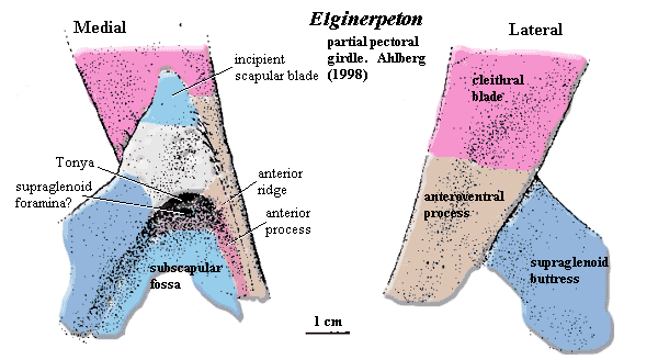
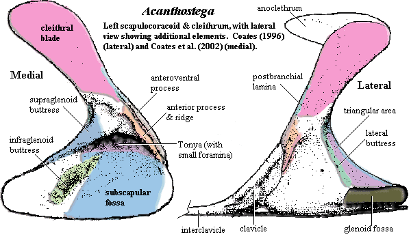
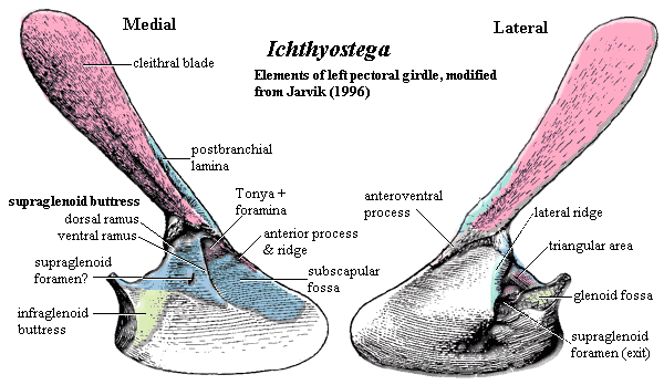
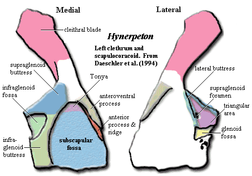
1. Anterior Process (of the scapulocoracoid): Like the anteroventral process of the cleithrum which covers it, this is a rather standard piece of equipment. It differs little from one critter to another. The anterior process runs down the anterior edge of the girdle, and marks the anterodorsal border of the subscapular fossa. For all or most of its length, it underlies the anteroventral process of the cleithrum. However, it is clearly visible as a more or less separate lamina. There is often a marked thickening, the anterior ridge, of the bone on the medial face, where it is not covered by the cleithrum. Anterior process of the scapulocoracoid = unnamed [A98] [D+94] [LC95] (not recovered in Tulerpeton), anterodorsal crest pars? [C96], coracoid crest pars? [J96],
2. Anterior Ridge: [D+94] notes an "anteromedial depression" on the scapulocoracoid anterior process, marking the limit of cleithral overlap. Coates [C96] points out that this is less accurately described as a "depression" than as the limit of a ridge on the exposed region of the scapulocoracoid where the anterior process is markedly thickened. We might add that Daeschler's [D+04] terminology also invites confusion with Jarvik's [J96] "anteromedial depression," which appears to be the posterior limit of the postbranchial lamina on the dorsal blade of the cleithrum. Anterior Ridge = unnamed "furrow" [A98], anterodorsal crest [C96], coracoid crest [J96], anteromedial depression [D+94], unnamed [LC95].
3. Anteroventral Process (of the cleithrum): This is a ramus of the cleithrum which descends the anterior margin of the girdle. Its rounded outer surface forms the articular condyle for attachment of the clavicle. Its inner surface overlaps the anterior process of the scapulocoracoid. The anteroventral process is remarkably similar in all pectoral girdles in this part of phylospace. The only real source of variation is the closeness of its relationship with the scapulocoracoid. In Elginerpeton, the two are tightly associated. Anteroventral process = anteroventral process of cleithrum [C96] [J96] [D+94], not named [LC95].
 4. Glenoid Fossa: The glenoid requires no
introduction. This is the joint on which the arm moves. It shows two
clear trends in the Late Devonian. It moves to successively more lateral
positions and (perhaps less clearly) becomes more strap-like. In Eusthenopteron,
it is offset medially from the medial face. In Panderichthys, it is
almost flush with the medial face. No part of the glenoid has been
recovered for Elginerpeton. In the 'stegas, the glenoid is
found at the level of the scapulocoracoid plate (Ichthyostega?) or a
little laterally (Acanthostega). In Hynerpeton and Tulerpeton
part of the glenoid is offset laterally a significant distance. This
is usually described in the literature as a change in orientation of the
glenoid. That is, the glenoid fossa is said to face progressively more
laterally. This manner of speaking confounds two completely different
trends and has caused all manner of difficulty. In at least Ichthyostega,
Tulerpeton, and probably Acanthostega, the fossa is curved,
perhaps in a rather complex manner. It is not possible to say in a few words
which direction the glenoid "faces." The current reconstructions
suggest, to a naïve observer (i.e., to us), that the angle is at least
bimodal. This might mean that the humerus was tightly restricted or
that it had two or more distinct articular modes. The geometry is complex;
and, without soft tissues, it is unreasonable to jump, hop, run, swim, or crawl
to any quick conclusions about locomotion.
4. Glenoid Fossa: The glenoid requires no
introduction. This is the joint on which the arm moves. It shows two
clear trends in the Late Devonian. It moves to successively more lateral
positions and (perhaps less clearly) becomes more strap-like. In Eusthenopteron,
it is offset medially from the medial face. In Panderichthys, it is
almost flush with the medial face. No part of the glenoid has been
recovered for Elginerpeton. In the 'stegas, the glenoid is
found at the level of the scapulocoracoid plate (Ichthyostega?) or a
little laterally (Acanthostega). In Hynerpeton and Tulerpeton
part of the glenoid is offset laterally a significant distance. This
is usually described in the literature as a change in orientation of the
glenoid. That is, the glenoid fossa is said to face progressively more
laterally. This manner of speaking confounds two completely different
trends and has caused all manner of difficulty. In at least Ichthyostega,
Tulerpeton, and probably Acanthostega, the fossa is curved,
perhaps in a rather complex manner. It is not possible to say in a few words
which direction the glenoid "faces." The current reconstructions
suggest, to a naïve observer (i.e., to us), that the angle is at least
bimodal. This might mean that the humerus was tightly restricted or
that it had two or more distinct articular modes. The geometry is complex;
and, without soft tissues, it is unreasonable to jump, hop, run, swim, or crawl
to any quick conclusions about locomotion.
5. Infraglenoid Buttress: this is the ridge or buttress on the medial face, located (roughly) opposite the glenoid fossa, or slightly ventral to that location. It forms the anterior and ventral margin of the infraglenoid fossa (if present) and the posterior margin of the subscapular fossa. It may be present in Elginerpeton, but that part of the pectoral girdle is largely unknown. The infraglenoid buttress sometimes appears as a ventral ramus of the supraglenoid buttress, as in Hynerpeton and Tulerpeton. However, the two supports are quite distinct in Acanthostega, if we are correct in finding (contrary to Coates) homology between the infraglenoid buttress and the "supraglenoid buttress" of [C96]. Our wavering confidence in this conclusion is bolstered by a consideration of Ichthyostega, which appears to have an intermediate condition. In both of the 'stegas the infraglenoid buttress plays a role in actually buttressing the glenoid, while the glenoid is entirely a structure of the lateral face in Tulerpeton and Hynerpeton. Infraglenoid buttress = infraglenoid buttress [LC95]; infraglenoid buttress pars [D+94]; supraglenoid buttress pars [C96], [J96].
6. Infraglenoid Fossa: This is a medium-sized depressed area on the posterior margin of the girdle, which is often thin and poorly preserved in early tetrapods (and so, very unlike the posterior margin of the things found in human girdles). It is bounded by the supraglenoid buttress dorsally and the infraglenoid buttress anteriorly and ventrally. The region is unknown in Elginerpeton.. In Acanthostega, Ichthyostega, and Tulerpeton, the infraglenoid fossa is probably absent, although it is conceivably homologous with the medial opening of the "Foramen A" of [C96] the "glenoid canal" of [J96], or an unlabelled foramen in [LC95]. Since these are all doubtful instances, this feature may turn out to be an autapomorphy of Hynerpeton, where it is plainly present. Infraglenoid fossa = infraglenoid fossa [D+94].
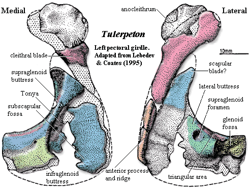 7. Lateral Buttress: We warned you about this one. This is the ridge running posterolaterally,
and, variably, also ventrally, on the lateral face. It forms the posterodorsal
border of the lateral face. Together with the supraglenoid buttress,
it defines the triangular area which constitutes the dorsal face. The lateral buttress usually
terminates above the glenoid fossa, at about the anterior limit of the
glenoid. In Elginerpeton, the lateral buttress is absent. The supraglenoid
buttress marks the entire dorsal margin, and there is no triangular area or
other distinct dorsal surface. By contrast, the lateral buttress is quite
distinct in Acanthostega, Hynerpeton, and Tulerpeton.
Tulerpeton differs from Acanthostega and Hynerpeton in that
the lateral buttress actually reaches and participates in the dorsal margin of
the glenoid. This is similar to the condition in Ichthyostega,
in which the lateral buttress projects sharply outward (i.e. laterally).
Coates [C96] has suggested that this is probably an artifact, and he seems to
have a strong argument. Lateral buttress = supraglenoid
buttress pars [C96], [D+94], [LC95]; anterior buttress [J96].
7. Lateral Buttress: We warned you about this one. This is the ridge running posterolaterally,
and, variably, also ventrally, on the lateral face. It forms the posterodorsal
border of the lateral face. Together with the supraglenoid buttress,
it defines the triangular area which constitutes the dorsal face. The lateral buttress usually
terminates above the glenoid fossa, at about the anterior limit of the
glenoid. In Elginerpeton, the lateral buttress is absent. The supraglenoid
buttress marks the entire dorsal margin, and there is no triangular area or
other distinct dorsal surface. By contrast, the lateral buttress is quite
distinct in Acanthostega, Hynerpeton, and Tulerpeton.
Tulerpeton differs from Acanthostega and Hynerpeton in that
the lateral buttress actually reaches and participates in the dorsal margin of
the glenoid. This is similar to the condition in Ichthyostega,
in which the lateral buttress projects sharply outward (i.e. laterally).
Coates [C96] has suggested that this is probably an artifact, and he seems to
have a strong argument. Lateral buttress = supraglenoid
buttress pars [C96], [D+94], [LC95]; anterior buttress [J96].
8. Postbranchial Lamina (of the cleithrum): In fish, the gills are braced at various points on the skull, frequently on the anterior edge of the cleithral blade, which then marks the posterior limit of the opercular chamber [CC91]. Even after the extrascapulars disappear and the pectoral girdle loosens from the skull, some proto- and early tetrapods retain a roughened anterior extension of the cleithral blade which presumably served this purpose. Its presence or absence has a peculiar phylogenetic distribution, and the lamina may not serve as a good proxy for gill breathing [A98]. See also [C96]. It is present in both of the 'stegas: strongly developed in Acanthostega, but only weakly in Ichthyostega. It is otherwise absent in this corner of phylospace, including the putatively primitive Elginerpeton. Postbranchial lamina = branchial lamina [A98] = postbranchial lamina [C96] [D+94] [LC95]; unnamed [J96].
9. Scapular Blade: In Late Paleozoic tetrapods, the scapulocoracoid develops a strong dorsal blade which functionally and morphologically replaces the dermal bone of the dorsal cleithral blade. Since none of our Devonian lot have a definitive scapular blade, it doesn't require much discussion. [LC95] figure a scapular blade in Tulerpeton. If present, it is small.
10. Subscapular Fossa: this is a large, somewhat triangular fossa which may occupy much of the medial face of the pectoral girdle. It becomes continuous with the ventral edge of the scapulocoracoid, which forms the base of the triangle. The two sides are subtended by the supraglenoid and infraglenoid buttresses, posteriorly, and the anteroventral process of the cleithrum (and/or anterior ridge of the scapulocoracoid) anteriorly. It is known to be present, by this name, in all five early tetrapods. However, there is an important ambiguity hidden in this apparent unanimity. It is sometimes unclear whether the reference is to the broad, normally flat, surface of the subscapular fossa or to Tonya (q.v.) at its apex. This may have caused confusion in the past, so we distinguish the two structures. Subscapular fossa = subscapular fossa [all].
11. Supraglenoid Buttress: this is the ridge or buttress running posterolaterally and ventrally on the medial face. It forms, in most cases, the posterodorsal border of the medial face. Together with the lateral buttress, it defines the dorsal triangular area. The supraglenoid buttress terminates (probably) at the posterior margin of the scapulocoracoid, but its full length is rarely preserved. The supraglenoid buttress also forms the posterodorsal margin of the subscapular fossa and the dorsal margin of the infraglenoid fossa (where present). In Elginerpeton, the supraglenoid buttress marks the entire dorsal margin, and there is no triangular area or other distinct dorsal surface. The supraglenoid buttress may appear continuous with the infraglenoid buttress, as in Hynerpeton and Tulerpeton. However, the two are quite distinct in Acanthostega. Ichthyostega appears to have an odd intermediate condition, also involving two distinct rami of the supraglenoid buttress. Supraglenoid buttress = infraglenoid buttress [A98], [D+94]; supraglenoid buttress [J96], supraglenoid buttress pars [C96], [LC95].
12. Supraglenoid foramen: We have made no attempt to account for the various holes in the girdle. With few exceptions, workers in this area wisely choose to refer to these foramina by letter designations. The soft tissues relating to the forelimb were being extensively rewired in the Late Devonian, and no one has yet been bold enough to publish their guesses about exactly what nerves and vessels were going where. One partial exception is the supraglenoid foramen. This hole is quite large in some species, and, like moths drawn to lighted windows, some workers have been unable to refrain from banging their heads against the glass. In fact, it is not completely clear that it is the same structure in all Devonian forms. This foramen was absent or not recovered in Elginerpeton. Ahlberg located a number of small foramina within Tonya, but he found no exits on the lateral face [A98]. In Acanthostega, there are a number of candidates, but Coates identifies none of them with the supraglenoid foramen. With respect to the other forms, we quote from [LC95: 313], stripped of citations and some examples, and with some changes in nomenclature:
The large, laterally directed supraglenoid foramen opens [laterally on the lateral buttress], and [medially] within the large subscapular fossa. This foramen resembles those of Proterogyrinus, Pholiderpeton, and Eoherpeton, and differs from the more numerous scapulocoracoid foramina of Acanthostega; its presence contrasts with the absence of any supraglenoid foramina in Greererpeton. Contrastingly, the restored pectoral girdle of Ichthyostega has a large supraglenoid foramen resembling that of Eusthenopteron. However, one of the authors (MIC, personal observation) disputes this: Ichthyostega has several small foramina [within in or just ventral to Tonya] like those of Acanthostega, and a moderately sized supraglenoid foramen within the triangular area, as found in Hynerpeton. Supraglenoid foramina, where known in [crown tetrapods] are also located within the triangular area (although near or [on the lateral buttress]), and are posterodorsally directed.
13. Tonya: There was no name for this structure, so we made one up. We were initially inclined to name it the "subscapular sulcus," but settled on Tonya for reasons which are absolutely none of your business. We were almost dissuaded at the last possible moment by a harsh look from our spouse, but decided we could probably slip this one by, and she'd never know the difference. Tonya is the apical portion of the subscapular fossa which is actually roofed over by scapulocoracoid bone. References in the literature to a "deep subscapular fossa" actually refer to the dorsoventral extent of Tonya. This has caused a certain amount of understandable confusion in the past. One of the plates in Jarvik's monograph [J96:167, pl. 45.2] appears to show a lamina of bone separating Tonya from the rest of the subscapular fossa. This is probably an artifact of preservation, but bears looking into. Tonya = subscapular fossa pars [all].
14. Triangular Area: The triangular area is the small dorsal (or posterodorsal) surface of the pectoral girdle. It is defined by the diverging arms of the supraglenoid buttress and the lateral buttress. The base is formed by the glenoid and/or the posterior margin of the girdle. The triangular area is absent in Elginerpeton, which lacks a lateral buttress. This area is rather beat-up in most specimens of Ichthyostega, but the triangular area is probably present. See pls. 45.4, 48.1 in [J96]. It is clearly marked in the other three animals under consideration. Triangular area = Triangular area [C96] [D+94] [LC95]; supraglenoid foramen (?!) [J96].
References: Ahlberg (1995) [A95]; Ahlberg (1998) [A98]; Ahlberg (2004) [A04]; Ahlberg & Clack (1998) [AC98]; Coates (1996) [C96]; Coates & Clack (1991) [CC91]; Coates et al. (2002) [C+02]; Coates et al. (2004) [C+04]; Daeschler et al. (1994) [D+94]; Jarvik (1996) [J96]; Lebedev & Coates (1995) [LC95].
 Elginerpetonidae:
Elginerpeton, Obruchevichthys.
Elginerpetonidae:
Elginerpeton, Obruchevichthys.
Range: Late Devonian (Frasnian); of Europe (Scotland) & Baltics.
Phylogeny: Elpistostegalia :: ((Metaxygnathus + Ventastega) + Tetrapoda) + * : Elginerpeton + Obruchevichthys.
Characters: dentary & anterior infradentaries strongly convex [A95$]; splenial and postsplenial fused [A95$]; deep furrow at dentary- infradentary suture; narrow, dorsal prearticular [A95]; fangs on the anterior coronoid separate from main tooth row (primitive) [A95].
Image: we think this is part of the right lower jaw of Eliginopteron in lingual view, showing a more or less complete anterior coronoid. Part of the edge of the Meckelian foramen can be seen at the right (posterior) end. The denticulated ridge of the prearticular is in the foreground, in front of the coronoid teeth. At the left, the coronoid toothrow appears disrupted -- probably by the offset roots of the anterior coronoid fangs. The medial lamina of the splenial can be seen sutured closely to the prearticular in the lower left quadrant.
References: Ahlberg (1995).
| Page Back | Unit Home | Page Top | Page Next |
checked ATW050624