| Palaeos |  |
Eureptilia |
| Vertebrates | Captorhinidae |
| Page Back | Unit Home | Unit Dendrogram | Unit References | Taxon Index | Page Next |
| Unit Back | Vertebrates Home | Vertebrate Dendrograms | Vertebrate References | Glossary | Unit Next |
|
Abreviated Dendrogram
AMNIOTA |--SYNAPSIDA `--+--ANAPSIDA | `--EUREPTILIA |--Coelostegus `--+--+--Thuringothyris `--+--Captorhinidae | `--Concordia | |--Concordia | `--+--Romeria | `--+--Captorhinus | `--Moradisaurinae `--+--Brouffia `--Romeriida |--Paleothyris `--+--Hylonomus `--+--Protorothyrididae `--DIAPSIDA |
Contents
Overview |
The Captorhinidae (in former times considered as 'stem reptiles') were one of the earliest and most primitive reptiles. They first occured in the Late Carboniferous of North America. In Upper Permian times they spread throughout the world but dissapeared from the North American fossil record, and finally they became extinct by the end of the Permian. Captorhinid fossils today are known from all continents with the exception of Antarctica and Australia. Captorhinids have an anapsid skull which is triangular in dorsal view and more or less heavily ornamented with a honeycomb-like pattern of ridges and pits, similar to that of numerous Late Paleozoic amphibians or, to a certain degree, modern crocodiles. Their body size ranges from that of a modern lizard up to that of a medium-sized modern alligator or the large Komodo Island monitor.
It has been mistakenly said that multiple tooth rows are a characteristic trait of captorhinids. This is not true and related to the fact that Captorhinus aguti, a representative with multiple rows of teeth, is the by far most common captorhinid. In reality captorhinids are divided into basal, rather small, lightly built forms with single rows of teeth, and into derived, larger forms with multiple rows of teeth. The most derived multiple-tooth-rowed forms form a clade referred to as subfamily Moradisaurinae. Cladistic analyses reveal that the evolution of multiple tooth rows took place at least three times independently within captorhinids: in Captorhinus, Captorhinikos and within the moradisaurines.
There is little doubt that the basal single-tooth-rowed forms were most likely feeding on insects and/or small vertebrates. The more robust built multiple-tooth-rowed forms seem to have fed on fibrous plants but an omnivorous diet or a diet consisting of hard-shelled invertebrates, such as clams or crabs, cannot be ruled out. Zidane080425 (from Palaeos org)
 Captorhinidae : Captorhinus.
Range: Permian of North America, Europe & East Africa.
Phylogeny: Eureptilia ::: (Protorothyrididae + Diapsida) + (Thuringothyris + * : Concordia + (Romeria + (Protocaptorhinus + (Rhiodenticulatus + (Saurorictus + (Captorhinus + (Captorhinikos + (Labidosaurus + Moradisaurinae))))))))
Characters: Robust skull; premaxilla downturned, with massive ventral base [MR05]; maxilla with narrow lateral exposure [MR05]; lacrimal suture with nasal usually mucg shorter than frontal suture [MR05]; lacrimal suture with nasal as long as frontal suture [MR05]; prefrontal usually wedged into posterior section of nasal [MR05]; pineal foramen on anterior part of parietals [MR05$]; supratemporals reduced or absent; tabular absent [MR05$]; jugal anterior process broad & blunt [MR05]; jugal with medial process [MR05]; large medial process of the jugal; single (lower) temporal fenestra; braincase supported by supraoccipital process; middle ear region open at least laterally and ventrally; ectopterygoid absent [MR05$]; external surface of lower jaw usually ornamented [MR05]; flat teeth; most have multiple rows of marginal teeth; number of maxillary teeth <25 [MR05$].
Links: Phylogeny and Classification of Amniotes; CAPTORHINIDA; Paleopedia - Cotylosaurs Palaeocritti - Eureptilia.
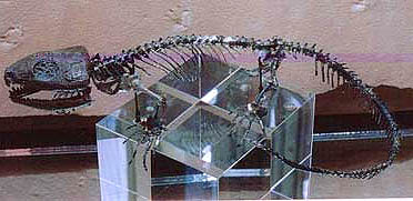
Image: Captorhinus aguti © 2000 Friends of the Geology Museum (Univ. of Wisconsin -- Madison), reproduced by permission. ATW010208.
References: Müller & Reisz (2005) [MR05]. ATW051015.
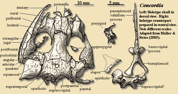
 Concordia cunninghami Müller & Reisz, 2005.
Range: Pennsylvanian (Gzhelian) of Kansas, USA. Known from two skulls.
Phylogeny: Captorhinidae : (Romeria + (Protocaptorhinus + (Rhiodenticulatus + (Saurorictus + (Captorhinus + (Captorhinikos + (Labidosaurus + Moradisaurinae))))))) + *.
Characters: premaxilla short, not overlapping lower jaw [MR05]; premaxilla without ventral curvature [MR05!]; premaxilla with 2-pronged nasal suture [MR05]; Type B septomaxilla, close to posterior border of naris [MR05]; maxilla slender & elongate [MR05]; maxilla dorsal lamina slight [MR05]; maxilla forming ventral border of naris, not extending to orbit [MR05$]; maxilla with well-developed horizontal lamina, expanded anteromedially [MR05]; nasals long, plate-like, rectangular, with slight expansion posteriorly [MR05]; lacrimal quite large, forming most of anterior & half of ventral orbital margin [MR05]; lacrimal dorsally expanded [MR05$]; dorsal & ventral lacrimal duct foramina on lacrimal in orbital margin [MR05]; lacrimal strongly interdigitating nasal suture [MR05]; lacrimal elongate contact with jugal [MR05]; prefrontal posteriorly elongate, almost reaching postfrontal & forming much of orbit dorsal margin [MR05]; prefrontal wide anterior to orbit, with sharp anterior tip wedged between nasal & lacrimal [MR05]; lacrimal suture with nasal as long as frontal suture [MR05]; prefrontal ventral lamina partially covered by lacrimal [MR05]; frontals rectangular, forming most of skull table above orbits [MR05]; frontals slightly expanded posteriorly, with serrated suture to parietals [MR05]; postfrontal triradiate, with sharp anterior point, and forming posterodorsdal orbit [MR05] [1]; postfrontal with posterior point inserted into parietal [MR05]; parietals short, much broader than frontals, & posteriorly embayed, creating 1 median and 2 lateral posterior processes [MR05!]; posterolateral process of parietal with slot for small, superficial supratemporal [MR05]; anterior of supratemporal on skull table, with ornament, but posterior occipital and unornamented [MR05]; parietal with extended occipital flange underlying paired, adjacent, semilunar postparietals on occiput [MR05]; tabulars absent [MR05]; jugal large, forming most of suborbital skull, with long preorbital process, short dorsal postorbital process, and flat temporal lamina [MR05]; jugal anterior tapers to a point [!] & inserts between maxilla & lacrimal [MR05] [2]; jugal dorsal process supports postorbital [MR05]; jugal temporal lamina extend below squamosal & probably contact quadratojugal [MR05]; jugal medial process absent [MR05]; jugal with distinct anterior and posterior ventral margins [MR05]; squamosal large, covering most of temporal region, bearing distinct occipital flange separated by vertical ridge [MR05]; quadrate broad in lateral view, with tall dorsal process [MR05]; basioccipital longer than broad, with anterolateral basal tubera [MR05]; basioccipital forming part of occipital condyle [MR05]; spraoccipital with well and "almost equally developed dorsolateral and ventrolateral processes surrounded by prominent, dorsally directed flanges. The semicircular canals are situated between the two projections." [MR05: 564] [3]; opisthotic stout and compact, with paroccipital process absent [4]; stapes short, with poorly developed shaft & large stapedial foramen [MR05!]; alar (posterior) basisphenoid projections probably contacted basisphenoid tubera [MR05]; parasphenoid completely fused to basisphenoid [MR05]; basipterygoid processes stout & directed anterolaterally [MR05]; parasphenoid cultriform process denticulate [MR05!]; vomer small [MR05]; pterygoids terminating anteriorly on midline between vomers [MR05]; interpterygoid vacuity present posteriorly [MR05]; pterygoid quadrate ramus with separate dorsal flange extending from basicranial articulation to dorsal process of quadrate, supporting elongate epipterygoid [MR05]; lower jaw largely unornamented [MR05]; lower jaw with long dorsal ridge for external jaw adductor [MR05!]; dentary quite straight, with abrupt curvature to symphysis [MR05]; long splenial present, not contributing to symphysis [MR05]; dentary makes up 75% length of jaw [MR05]; retroarticular process absent [MR05!]; articular small and lens-like [MR05]; teeth generally small, homodont, pointed & recurved distally [MR05]; 5 small, pointed, premaxillary teeth [MR05]; caniniforms absent, but 4 anterior maxillary teeth somewhat elongate, others shortening posteriorly [MR05!]; single maxillary tooth row with 18 teeth [MR05]; probably 17 dentary teeth [MR05]; 2 vomerine tooth rows, with medial row continuing pterygoid denticles [MR05]; vomerine lateral denticles autapomorphic [MR05$].
Notes: "!" indicates feature unique among captorhinids, but likely plesiomorphic for Eureptilia. [1] The postfrontal in the referred specimen has a different shape. It is probably simply broken, but the bone labelled "postorbital" in the referred specimen has the correct shape for the postfrontal, and might have been dislodged by a fragment (labelled "postfrontal"), which could, in turn, be a piece of the thoroughly splintered epipterygoid. These skulls were both recovered from split shales. The fossil side was then embeded in resin and apparantly prepared from the opposite (rock) side. This, the authors note, created some difficulties in identifying pieces deeply embedded in the resin, such as the circumorbital bones of the referred specimen. [2] One problem with getting too artistic is that it can obscure details. Our attempts to gussy up the figure from [MR05] obscure this insertion, which is quite clear in the original image. [3] Note that the semicircular canals, i.e. normally the guts of the otic capsule, are described as lying dorsal to the supraoccipital. Ummm. We have concerns about this reconstruction of the occiput and posterior braincase. After struggling with it for some hours, we've decided that we lack sufficient expertise to make any comments at all, beyond this vaguely dissatisfied grunting noise. We therefore grunt, and readers may make of that what they will. [4] The paroccipital process is said to be absent. However, observe the opisthotic illustrated on the left side of the holotype (dorsal view) image here. It shows an unprecedented ventromedially directed process. Ummm. See footnote 3 for explanation of grunting noise. ATW051016
Comments: Concordia cunninghami is the oldest known and most basal captorhinid so far and the only recognized species of the genus. It is known from two almost complete skulls coming from Upper Pennsylvanian rocks of the famous Hamilton Quarry in Greenwood County, Kansas.
C. cunninghami not surprisingly shows many features that are typical for basal eureptiles but are not present in more derived captorhinids. The premaxilla is small and seems not to have formed a down-curved "beak" as it is seen in almost all of the remainder of the captorhinids. The lacrimal is dorsoventrally expanded and forms the main portion of the lateral wall of the snout which is similar to the condition in Rhiodenticulatus. The skull table lacks the tabular bone, a feature considered as synapomorphy among captorhinids. The supratemporal is small as well and points, in dorsal view, anteromedially into the posterolateral corner of the parietal as seen in another basalmost captorhinid, Romeria, or in the "Protorothyridid" Protorothyris. The same applies for the posterior margin of the skull roof which is embayed bilaterally in contrast to the pattern present in more derived captorhinids where this margin posesses a single median embayment. Like in Romeria and Protocaptorhinus the parietal foramen is very large compared to the overall size of the skull.
Maxilla and dentary are lined with a single row of as much as 18 small, pointed teeth. The palatal bones and the basisphenoid (a ventral element of the braincase) are covered with numerous denticles. A unique feature of C. cunninghami is that the vomer not only exhibits medial rows of denticles but, in addition, has also lateral ones. An important captorhinid apomorphy, the reduction of the ectopterygoid and replacement of that bone by the adjacent ones, seems to be present in C. cunninghami where it is obviously replaced by the transverse process of the pterygoid. In Captorhinus, however, it is rather a medial process of the jugal that occupies the position of the ectopterygoid. A remarkable occipital feature is that the opisthotic seems not to have the lateral projection which usually contacts the cheek bones, known as paroccipital process. This process is a characteristic trait in all basal amniotes as well as in more modern reptiles, such as squamates.
The oldest known eureptile and one of the closest relatives of captorhinids is the "Protorothyridid" Hylonomus from the Upper Pennsylvanian of Nova Scotia. Thus, although still geologically younger, the discovery of C. cunninghami considerably shortens the ghost lineage of the captorhinids.
Some Facts
Family: Captorhinidae
Etymology of genus: "unity, agreement, harmony", refers to the fact that the occurrence of a captorhinid in Upper Pennsylvanian strata confirms the long-held assumption that captorhinids must have existed as early as in Late Carboniferous times
Etymology of species: named after the paleontologist Christopher R. Cunningham
Paleography: Cherokee Basin, northwestern Pangaea
Locality: Greenwood County, Kansas, USA
Horizon: Calhouns Shale (Shawnee Group)
Stratigraphic Range: Upper Pennsylvanian (Late Carboniferous): Virgilian
Zidane 080526 (Palaeos org)
References: Müller & Reisz (2005) [MR05].
| Page Back | Unit Home | Page Top | Page Next |
checked ATW050509
Using this material. All material by ATW is public domain and may be freely used in any way (also any material jointly written by ATW and MAK). All material by MAK is licensed Creative Commons Attribution License Version 3.0, and may be freely used provided acknowedgement is given. All Wikipedia material is either Gnu Open Source or Creative Commons (see original Wikipedia page for details). Other graphics are copyright their respective owners