| Glossary | ||
| The Vertebrates | H-I |
| Vertebrates Home | Vertebrate | Vertebrate | Bones | Time |
For most phrases beginning with directional words, e.g. "posterior," "dorsal," "external," etc., or some generic anatomical terms, e.g., "vena," look under the next word in the phrase. However, note that this convention is not used with complete consistency in this Glossary.
Habib Rahi FormationMiddle Eocene Lutetian) of Pakistan. Marine. Gingerich et al. (2001).
Haeckel's Law ontogeny recapitulates phylogeny. Neither true nor false, Haeckel's Law remains an important tool in understanding and analyzing phylogenetic problems.
Hair cell a mechanosensory cell characterized by the presence of microvilli. When the microvilli are deformed by mechanical pressure or vibration, the cell increases or decreases the rate at which it sends an electrical signal to the nerve cell with which it is coupled. Hair cells frequently are highly directional. If the villi are bent on one direction, the rate of discharge increases. In the opposite direction, the rate decreases. If the villi are deformed at right angles to this axis, there may be no change in the signal. Hair cells are the basic cellular unit involved in hearing, in the labyrinth (balance organ), and in the lateral line system of fish.
Halite a mineral composed mainly of table salt. NaCl.
Hallux digit 1 of the foot in birds, normally reversed to point posteriorly, presumably as an adaptation for perching, rather than out of sheer perversity. Digits are counted from medial to lateral, so this is the big toe.
Hamular hook-shaped.
Harding Sandstone Late Ordovician (Sandbian) of North America (Colorado). Vertebrate microfossils, including Astraspidae, Eriptychius, and possibly thelodonts and sharks. Sansom et al. (1996).
Harudi Formation Middle Eocene Lutetian) of far western India (Gujarat). Bajpai & Thewissen (1998). Remingtonocetus
Hauterivian an age of the Early Cretaceous about 132-127 Mya.
Haversian Canal the central canal in an osteon. The Haversian system refers to the organization of advanced vertebrate bones characterized by osteon units.
Haymana Basin Late Cretaceous to Pliocene of Turkey (Central Anatolia). Forearc accretionary basin sediments accumulated during closure of Tethys Sea along its Northern margin. Mammals.
Heath Formation Early Carboniferous of Montana, part of the Big Snowy Group. Mostly near-shore marine, near a very arid terrestrial environment (about 10° S paleolatitude) with little or no fresh water contribution. Includes the Bear Gulch Limestone. May extend geographically south into the Dakotas and temporally into the Late Carboniferous.
Hell Creek Formation Late Cretaceous (Maastrichtian) of Montana. Dinosaurs, birds, multituberculates, crocs, turtles & champsosaurs.
Hemapophysis The second element in each half of a hemal arch, corresponding to the sternal part of a rib. In Paleozoic chondrichthyans, hemapophyses are frequently present in the tail and for an important part of the attachments for basals and radial elements forming the ventral tail lobe. See Orodontida.
Hemochorial placenta type of a chorioallantoic placenta in which the villi are in direct contact with maternal blood supply.
Hendricks Formation Early Silurian (Llandovery) of North America (Wisconsin), Burnt Bluff Group. Coral-stromatoporoid intertidal environment found as wackestone. Thelodont scales. Turner et al. (1999).
Hepatic relating to the liver.
Hepatic diverticulum an evagination of the embryonic gut i.e. the endoderm) which invades the surrounding coelom and, specifically, the transverse septum. The diverticulum develops into the liver and tastes revolting, although it is quite good for you and may remain fairly diverting with spinach and crisp bacon, accompanied by a reasonably mature Merlot.
Heterocercal same as epicercal, according to some sources, or (perhaps better) referring to the condition in which the caudal fin is asymmetric as either epicercal or hypocercal. See PPT Slide.
Heterodactyl a specialized digital configuration in birds in which both digits 1 and 2 are reversed.
Heterodont bearing teeth of more than one sort. The shapes may change progressively or abruptly. The term is usually used to imply more than a change in size. Humans have heterodont dentitions, consisting of incisors, a canine, premolars, and molars.
Heterothermy metabolic temperature regulation, but regulation is imprecise or variable.
Hirnantian Age third and last age of the Late Ordovician, 446-444 Mya. See Hirnantian.
Histo- Greek root meaning tissue. Supposedly derived from a word meaning web or loom.
Histogenesis development of tissues; differentiation of tissue types in development.
Histology study of the structure of biological tissues.
Holocephalic (or holocephalous) of ribs, having one head. That is, having a single articulation with the vertebrae, as opposed to dichocephalic or dichocephalous, having two articulations.
Holospondyly a condition in which all of the vertebral elements are fused. Opposite of aspidospondyly. Sometime used in a more restricted sense to mean the condition in which there is only one centrum per vertebra, rather than separate pleurocentra and intercentra.
Holostylic a type of jaw suspension in which the palatoquadrate is fused to the braincase.
Holotype A single specimen designated as the name-bearing type of a species or subspecies when it was established, or the single specimen on which such a taxon was based when no type was specified.
Homeobox The DNA sequence that codes the homeodomain. See homeodomain.
Homeodomain "the homeobox [sic] is a 60-amino acid helix-turn-helix motif that acts as a DNA-binding domain. Homeobox-containing genes are usually involved in developmental processes, such as embryonic patterning, organogenesis, and/or cell differentiation." Lanctôt, et al. (1999: 1416). This is succinct and (with one glaring exception) technically correct, but a horrible definition for beginners. To add at least a little background, note the following: (1) this is actually the definition of the homeodomain -- one of many known protein structures that bind DNA. The "homeobox" is the DNA sequence which codes for this protein fragment. The current generation of molecular biologists is incredibly sloppy in failing to distinguish between (a) the gene (including all regulatory sequences), (b) the DNA coding sequence, (c) the RNA transcript including exons, introns, etc.), (d) the ultimate mRNA, (d) the translated polypeptide, and (e) the final functional protein (with subunits, modified side-chains, etc.). These are critical differences, and it is important that one knows exactly which one is meant. Actually, the homeodomain is none of the above. It is a "motif," a sequence of amino acids found, with minor variations, in many proteins. (2) The "helix" referred to is the well-known alpha helix formed in any number of protein sequences. A simplified explanation can be found at PPS 96' - Alpha-Helix Geometry Part. 2. (3) The homeodomain seems to bind DNA without much specificity except that it requires a TAAT sequence [interestingly, and not coincidentally, RNA polymerases use the sequence TAATAA to locate the transcription initiation site]. (4) Homeodomain- containing proteins also contain a third alpha- helical segment which supplies the detailed sequence specificity. The homeodomain segment simply aligns the protein in the groove of the DNA double helix so that the protein's "recognition helix" is brought into contact with the base pairs of the DNA. The recognition helix is structured so that it binds only to specific sequences of DNA bases.
Homocercal of caudal fins, having the lower and upper fin lobes of approximately the same size and shape.
Homodont All teeth in the jaws are of the same shape, although they may be of slightly different proportions and quite different sizes.
Homology the relationship between structures in different organisms which are united by modification of the same structure, gene or set of genes of a common ancestor. Examples: whale caudal fins, lizard tails, bird pygostyles and the human coccyx are homologues. As contrasted with analogy, the relationship of two structures, genes or gene sets that are related by present structural or functional similarity without regard to phylogeny. Dolphins and ichthyosaurs share many analogous functional units, although their last common ancestor was likely a terrestrial proto-lizard. Both factors may be acting at once. Thus, prosauropods and sauropods had a sauropodomorph last common ancestor in the mid-Triassic. They diverged, but both developed quadrupedal gigantism of a similar type convergently, presumably in response to similar ecological pressures.
The strict Patterson formalism for homology requires that possible homologues satisfy the tests of similarity, congruence and conjunction. These tests can be summarized as follows: Similarity: Each homologue must have the same 1:1 topographical relationship with other structures. In practice, we may add "or a really good developmental explanation of why it differs." Congruence: The presence of the homologous character must be congruent with the cladogram. That is, the homology must be a synapomorphy of some clade. Conjunction: If two structures are homologous, then both can never appear in the same organism. See, e.g., Wittmer 1995).
Homoplasy a character which is shared by two taxa, but not by their common ancestor. That is, it arose independently in the two lineages and represents a convergence.
Honda Group Middle Miocene of Columbia. Cifelli & Villarroel (1997).
Hornerstown Formation Late Cretaceous (Maastrichtian) or Early Paleocene of New Jersey, USA. The "Main Fossiliferous Layer" is apparently terminal Maastrichtian. Glauconite greensands. Livezey 1997); Olson & Parris (1987). Anatalavis
Horseshoe Canyon Formation Late Cretaceous (early Maastrichtian) of Alberta, Canada. Holtz 2001a).
Huayquerian Age South American Land Mammal Age corresponding to the Late Miocene.
Humerus Nature's way of connecting the forearm to the shoulder. Sadly, this is an incredibly complicated bone and has been given its own page. See Humerus.
Hunter-Schreger bands in mammalian dentition, the enamel layer is acellular, being made up of closely packed, hydroxyapatite crystallites (? how does this differ from a crystal?) laid down by ameloblast cells in the form of enamel "prisms" or rods, which are arranged more or less at 90° to the surface of the tooth. It's the "more or less" that leads to the formation of bands. Alternating layers of enamel rods make roughly equal and opposite departures from this angle. The crystallites are literally "prisms" in their effect on light. As a result, the different layers show up as alternating light and dark bands under polarized light. These are the Hunter-Schreger bands. One might speculate that this is also why clean, healthy teeth literally "gleam" under appropriate circumstances (such as under the powerfully suggestive influence of low lighting and pheromones). See An Atlas and Practical Guide to Histology.
Hyaline cartilage cartilage with a clear translucent matrix; found primarily on the ends of ribs and on the trachea. Introduction to the skeletal system; lab7 photos
Hydroxyapatite the form of apatite in which the monovalent ion is the hydroxyl (OH ¨ ) ion. See long-winded explanation at apatite.
Hyoid Arch the second hypothetical ancestral gill arch, from which derives the hyomandibula and other elements of the splanchnocranium. See Gill Arches.
Hyodont of teeth, not ankylosed to the jaw.
Hyomandibula (= hyomandibular) the upper (epal) main element of the hyoid arch. The same bone as the stapes, columella, and epihyal. See Gill Arches.
Hyostylic a form of jaw suspension (e.g. in elasmobranchs and teleostomes) in which the upper jaw loses any major direct connection with the braincase and the upper and lower jaws are supported solely by the hyomandibula. Introduction to the skeletal system.
Hypantrum see hyposphene-hypantrum
Hypapophysis a ventral "spine" or keel on a vertebra. See Axial Skeleton.
Hyperphalangy the condition of having numerous additional phalanges (toe or finger bones).
Hypoblast see Gastrulation.
Hypobranchial in fishes, small gill arch elements which forms joints between the main ventral elements, the ceratobranchials, and the basibranchials, which are usually fused or tightly bound to the ventral aspect of the pharynx (throat).
Hypocaudal lobe the part of the caudal fin below the notochord.
Hypocercal caudal fin structure in which the notochord or vertebral centra extend only into the lower fin lobe.
Hypochordal [1] of a fin, same as hypocercal, but refers expressly to the notochord. [2] of a fin lobe, the caudal fin lobe below the notochord.
Hypocleidium an enlarged, flattened ventral area of the furcula at the distal end, formed by the fused clavicles. The pectoralis muscle attaches, in part, to the hypocleidium (if present).
Hypocone The main cone on the talon (distolingual extension) of a mammalian (tribosphenic) upper molar. See Molars, or figure at mesostyle.
Hypoconid in mammalian dentition, on the lower molars, the buccal main cusp of the talonid. In a quadrate-type molar, this would be the buccodistal main cusp. I understand there is an alternate system of nomenclature in which the terms hypoconid and entoconid are reversed and the hypoconid is the lingual main cusp of the talonid, i.e. what is referred to in these Notes as the entoconid. See Teeth, diastema lengths.
Hypoconule in mammalian dentition. Apparently, this is the same as the metaconule, that is, a small cusp on an upper molar lying near the line between the protocone (the lingual main cusp) and the metacone the distal main cusp). If the line is marked by a ridge, the ridge is the postprotocrista. By "main cusp," I mean one of the large cusps defining the vertices of the trigon. See figure at mesostyle.
Hypoconulid in mammalian dentition, a cuspule (accessory cusp) usually located near the distal (posterior) edge of a lower molar. See Molars.
Hypoglossal foramen the foramen for the exit of the hypoglossal nerve, usually located near the base of the occipital condyle. The hypoglossal nerve may also exit as part of the jugular or vagus foramen, or (from the scala tympani) in or near the metotic foramen.
Hypoglossal nerve The XIIth cranial nerve, with both sensory & motor fibers. Enervates , e.g., base of tongue.
Hypohyal in fishes, the hypohyal is a ventral element of the hyoid arch which links the ceratohyal and the basihyal. The gill arches have two basic parts, dorsal and ventral. Each part is associated with a main gill arch segment, the dorsal) epal and (ventral) ceratal segments, respectively. Thus, each of the branchial arches, the arches which actually function as respiratory arches in fishes, has an epibranchial and a ceratobranchial. The hyoid arch is a an additional arch anterior to the first branchial arch. The hyoid is often involved in respiration, but its primary functions are related to jaw support and extension. The ceratal and epal segments of this arch are called the ceratohyal and the hyomandibula, respectively. Most gnathostomes also have some kind of ventral extension of the gill arches, a basal component, and these usually lie flat along the ventral "throat" of the fish. The basal elements of adjacent arches are often closely interlocked, or even fused. The basal element of the hyoid arch, the basihyal, is no exception. The ceratal segment is typically long and relatively rigid and partially vertical, while the basal elements are typically fused and/or tightly bound to the base of the gullet in a horizontal orientation. Keeping these elements in articulation as they move in different planes requires a complex joint. This joint is normally supplied by a small element of complex shape. This element is the hypobranchial or, in the case of the hyoid arch, the hypohyal. See Hypohyal.
Hypolophid in mammalian dentition, a cutting edge running generally along the buccal side of the talonid of a lower lophodont molar. See image at lophodont and Molars.
Hypomere See Early Development Terms.
Hyposphene-hypantrum articulation "In many primitive theropods (the Triassic Coelophysis, for example) and allosaurs the pre-zygapophyses surfaces have two distinct articular planes: 1) the primary zygapophysis, a surface facing mostly upward and inward; and 2) a hypantrum, a surface facing mostly inward and downward. The right and left hypantra enclose a narrow triangular space below and between the primary joint surface of the zygapophyses. Into this space fits the hyposphene on the posterior of the preceding vertebra. Hyposphene-hypantra joints increase the backbone rigidity - the vertebra can flex side-to-side and up-and-down but cannot twist at all...." Megalosaurid Dinosaurs from the Late Jurassic [site defunct] [Siegwarth, JD, RA Lindbeck, PD Redman, EH Southwell, & RT Bakker (1999?)] See figure at right and also figure at zygapohysis.
Hypostyle A stylar cusp near the buccomesial corner of an upper tribosphenic molar. See figure at mesostyle.
Hypotarsus in birds, a ridge or process located on the posterior side of the tarsometatarsus, near the proximal end. It may be derived from the calcaneum. The hypotarsus is the attachment point for the posterior cruciate ligament, which originates on the posterodorsal part of the intercondylar groove of the femur (just about opposite the patellar sulcus). See Ch 1-5. Image adapted from Topographische Anatomie der Hintergliedmaße beim Habicht ( ....
Hypsodont having teeth with high crowns (cusps). Hypsodont dentition is associated with a diet of abrasive foods. Hypsodont teeth are frequently ever-growing. See The Diversity of Cheek Teeth at Animal Diversity Web. Opposite of brachydont.
Hypural in fish anatomy, one of the flattened, fused bony hemal spines fanning out at the base of the caudal fin and bearing caudal rays. Amia has 10 hypurals, Salmo 7 but in most teleosts the number is reduced or fused into a single structure, the urostyle. (from the Dictionary of Ichthyology). See image at urodermal.
Hypurapophysis see parhypural.
I/i in mammalian dentition either an upper (I) or lower (i) incisor. In English grammar, the nominative case of the first person singular pronoun (I) or, in the case of the poet e.e. cummings or one of his many imitators, (i).
Iliac pertaining to the ilium
Iliac peduncle the ilium often has two peduncles (ventral processes) which come down on either side of the acetabulum to form the sides of the acetabulum and meet the pubis and ischium. These are referred to as the pubic peduncle and the ischiac peduncle (or, sometimes, pedicle). By analogy, if the pubis or ischium have dorsal process which come up to meet the ilium, this structure is referred to as an iliac peduncle.
Iliofemoralis The m. iliofemoralis is a muscle of variable importance which, as the name implies, originates on the ilium and inserts on the lateral femur. Hutchinson & Gatesy (2000). In turtles, the muscle is rather massive and inserts on an almost unique structure, the trochanter major. A similar, conceivably homologous, structure is found in some pachypleurosaurs. Carroll & Gaskill (1985). In birds, the anatomy is complicated by terminology. The reptile iliofemoralis is the m. iliotrochantericus caudalis and the iliofemoralis externus of birds, while the muscle called the "iliofemoralis internus" is the homologue of one of the several reptile mm. puboischiofemoralis. Carrano & Hutchinson (2002). Primitively, the origin is above the acetabulum, but it is moved far forward in most archosasurs, where it starts on and under the anterior iliac blade (id.; Currie & Zhao, 1993) and inserts on the internal trochanter (Hutchinson, 2001a). It generally acts to raise or abduct the femur. However, in cynodonts, due to the different geometry of the femur, it came to act as a retractor (Carroll, 1988), with a tendon running through a notch in the supraacetabular buttress (Surkov et al. 2005). Eventually (in mammals) it was absorbed into the gluteal musculature. Carroll (1988).

Iliofibularis tubercle the m. iliofibularis is a muscle originating high on the ilium, posterior to the acetabulum, often involved in flexing the lower leg. It inserts on the fibula, near the proximal end. The point of insertion may be marked only by a rugose (roughened) patch. However, it may also insertion a more or less prominent tubercle, the iliofibularis tubercle, or even an iliofibularis trochanter. This seems to be the same thing as the anterior trochanter of the fibula.
Iliotrochantericus a deep, relatively small muscle of the pelvis, normally (in birds) with separate cranial & caudal origins on the lateral face of the ilium. Both heads insert on the femoral trochanter and presumably act as protractors and/or to stabilize the femur.
Ilium L. ilium = the flank and the iliac bone. The dorsal bone of the three bones forming the pelvis. The ilium supports the sacrum. Originally so called because the small intestines are largely supported by this bone, and the old term for the small intestines was ilia (plural of ilium). See figure at antitrochanter.
Imbricatingoverlapping. Sometimes used simply to indicate a complex pattern made up of small parts.
Impedance acoustic impedance is the resistance of a medium to the propagation of sound, and is largely a function of density and the speed of sound in the medium. In a slightly different sense, it is a measure of the resistance of an interface to the propagation of sound from one medium to the next. See The Ear.
Incisive foramen a foramen in the palatal process of the premaxilla just posterior to the incisors.
Incisor L. incidere = to cut into; from in = in, and caedere = to cut. Applied to the cutting teeth of the anterior jaw.
Incisure (= incisura) [L incidere to cut into] a cut, notch, or incision; a general term for an indentation or depression.
Incrassate of teeth, labiolingually expanded.
Incudomalleal joint the joint between the malleus and incus in the middle ear of mammals. See The Incus.
Incus L. incus (incudis) = anvil. The quadrate, when it is exapted as an auditory ossicle in mammals. See The Incus.
IncumbentLying, leaning, or resting on something else: incumbent rock strata.
Induan Age The first Age of the Early Triassic, about 248-245 Mya. Probable age of the Lystrosaurus Zone of the Karoo and elsewhere. See Induan
Induced drag no, this has nothing to do with cross-dressing. Induced drag is drag created incident to the production of lift. See Induced Drag.
Inferognathal (adj) relating to the lower jaw; (n) bone "tooth" plate(s) on the lower jaw of placoderms.
Infradentary a serially homologous group of bones ventral to the dentary. Basally, they are long bones running generally anterior to posterior, but at an angle to the line of the dentary. In Osteolepiformes and, progressively, in tetrapods they obey Williston's Law very nicely. That is the originally serially homologous group becomes fewer in number and more specialized. The most dorsal member (sometimes not considered an infradentary) is the surangular. This is followed by the angular and the splenials (if any). See image at Surangular.
Infrahemal autogenous spines distal to the hemal arches.
Inframeckelian Foramen a small foramen on the inner surface of the lower jaw, normally located on the suture between the splenial and prearticular and (as you may have guessed) below the level of the Meckelian cartilage.

InfundibulumAny of various funnel-shaped bodily passages, openings, structures, or parts, especially: a. The stalk of the pituitary gland. b. The calyx of a kidney. c. The ovarian opening of a fallopian tube. With respect to dentition, it refers to an infolded area on the occlusal surface.
Inguinal L. inguen = groin.
Innominate In mammals, the hip bone, consisting of three consolidated bones, the ilium, ischium, and pubis.
Insertion L. in = in, and serere = to put. Hence, the point of attachment of a muscle in the more movable of the two structures which it joins.
Integument skin. A typical arrangement of dermis is shown in the figure. The outer, usually acellular, layer may be composed of scales, cuticular material, mucous, etc. The columnar epithelial layer may contain invaginations forming numerous structures including hair or feather roots, endocrine or exocrine glands, sensory structures, tattoos, and so on. The epithelium is an ectodermal structure, and its inner boundary is almost always marked by a fibrous basement layer (which has additional microstructure not discussed here). The dermis is formed from mesenchyme and/or mesoderm. The dermis produces and supported the production of dermal bones and osteoderms. The hypoderm is a transitional layer of loosely linked connective tissue and subcutaneous (adipose) fat. Finally, the integument is generally bound, as a unit, to the body wall musculature. For development, see epidermis and dermis entries.
Intercalarium an occipital bone of actinopterygian fishes and one of the Weberian ossicles in Otophysi. See also image at claustrum.
Intercentrum the vertebral centrum associated with the interneural arches, if present. The intercentrum is formed in the center of each myomere and is thus formed by a single myomere, as opposed to the pleurocentrum, which is formed between two adjacent myomeres.
Interdorsal "There are primitively two pairs of [metamerically arranged endoskeletal] elements in each metamere and on each side [of the notochord]: the interdorsals and basidorsals. In the gnathostomes, there are two additional pairs ventrally to the notochord: the interventrals and basiventrals. These elements are called arcualia and can fuse to a notochordal calcification, the centrum. The ensemble of the arcualia + centrum is the vertebra, and the ensemble of the vertebrae is the vertebral column." See Vertebrata Phillipe Janvier).
Intermediate mesoderm See Early Development Terms.
Intermedium one of the proximal carpal bones of the wrist. See figure at carpus.
Internal ____ for most phrases beginning with directional words, e.g. "posterior," "dorsal," "external," etc., or some generic anatomical terms, e.g., "vena," look under the next word in the phrase. However, note that this convention is not used with complete consistency in this Glossary.
Interneural arch In the earliest vertebrates, it is supposed that each embryonic segment (myomere) of the spine contained two dorsal arches -- or actually one arch and two half-arches. One arch developed in the middle of the segment. At each end of the segment another arch was formed which fused with the arch forming at the end of the adjoining segment. With breathtaking illogic, the arches in the middle of each segment are referred to as interneural arches. The arches formed between two segments are then, by default, the neural arches.
Interpterygoid vacuity an open palate. See image. That is, a palate with open space between the pterygoids and the anterior ("cultriform") process of the parasphenoid. Not to be confused with the choanae, which are the posterior nasal openings normally located between the anterior marginal bones and the vomers (as in the image), or the suborbital fenestra (not shown), a second pair of marginal fenestrae found in lepidosauromorphs and related forms.

Interspinous notch a notch formed by anterior (or posterior) processes from the bases of the two halves of the neural spine where they meet at the midline. See image from Sill 1974).
Intertemporal bar a bar of bone separating the upper and lower temporal fenestrae in reptiles.
Intertrochanteric fossa a fossa or depression usually located directly under the head of the femur, for insertion of an important portion of the PIFE adductor musculature or its homologue.
Intracranial joint in Sarcopterygii, a complete transverse division of the braincase into anterior and posterior halves. The division runs between the basisphenoid & basioccipital ventrally and immediately anterior to the otic capsule dorsally.See image at otic shelf.
Intraprezygapophyseal lamina reinforcing ridge bone ridge in the vertebrae (normally, of sauropods) connecting the prezygapophyses. See image at centrodiapophyseal lamina.
Involucrum usually used in medical jargon to mean a sheath of replacement bone grown over dead or diseased bone, as in osteomyelitis. Used more generally to mean any bone sheath and, in particular, a sheath formed as an extension of the tympanic into the middle ear. The term is most frequently used by whale people who are specifically referring to a pachyostosis of the medial rim of the bulla which is enormously expanded, even in quite early whales. Luo 1998).
Iren Dabasu Formation Late Cretaceous, but very uncertainly dated, of North China (Inner Mongolia). Holtz 2001a) The formation directly overlies a slate floor of Cambrian age and is overlain by Eocene lake deposits of the Arshanto Formation. The Iren Dabasu sediments are clay with a few carbonate lenses. Vertebrates are found in the upper third of the formation, probably from a lowland lake depositional environment. Recovered vertebrate fauna include advanced theropods, an ankylosaur, turtles, fish and crocodiles. Mader & Bradley (1989).
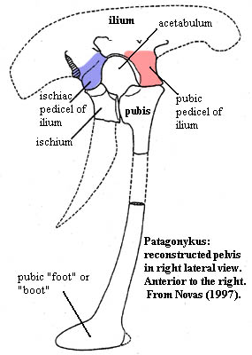
Ischiac pedicle of the ilium, the posteroventral extension of the ilium below the iliac blades which forms the posterior margin of the acetabulum and contacts the ischium. See blue area in figure at right. See also pubic peduncle.
Ischial peduncle (of ilium) same as ischiac pedicle or ischial pedicle.
Ischigualasto Formation Late Triassic (Carnian) of South America (Argentina). One of the great vertebrate fossil beds of the world and the source of countless important archosaur specimens of all flavors.
Ischiumthe posteroventral member of the three bones forming the pelvis. See figure at antitrochanter.
Isopedin the material forming a basal layer of laminal, dermal bone in paleozoic fish scales -- ossified, or largely ossified, layers of connective tissue. Also apparently used to describe any laminar dermal bone.
Itaboraí Formation Late Cretaceous or Early Paleocene of Brazil. Mainly limestone with sandy marl fissure and channel fills of Late Paleocene (Riochican) age. Didolodonts and litopterns in fissure fills. Cifelli 1983).
checked ATW031004