| Glossary | ||
| The Vertebrates | E-En |
| Vertebrates Home | Vertebrate | Vertebrate | Bones | Timescale |
For most phrases beginning with directional words, e.g. "posterior," "dorsal," "external," etc., or some generic anatomical terms, e.g., "vena," look under the next word in the phrase. However, note that this convention is not used with complete consistency in this Glossary.
Ec abbreviation for the Eocene.
Eccrine glands L. ex + crinis (hair). Sweat glands under sympathetic nervous system control, primarily involved in thermoregulation. As the name suggests, eccrine glands are not associated with hair follicles. Compare apocrine gland.
Ectal outer or exterior. In particular the outer (more peripheral) contact between the calcaneum and astragalus in mammals. See figure at Protocetidae.
Ectepicondylar foramen see Humerus.
Ectepicondyle a process on the distal humerus (upper arm bone) on the outer or ulnar side. It is associated with attachment of the extensor muscles of the lower forelimb. See Humerus.
Ectocuneiform the cuneiform is one of the bones of the ankle (a distal tarsal). It articulates proximally with the navicular and distally with metatrsals III and/or IV (the latter particularly in ungulates and some marsupials). In some types, the cuneiform is found as two or three bones, in which case the more anterior ("dorsal" in dorsal/plantar terminology) is the ectocuneiform, while the more posterior or plantar bones are the mesocuneiform and entocuneiform. Image: Brandoni et al. (2004).
Ectoderm One of the three primordial germ layers of all triploblast animals. Ectoderm may be the "original" tissue of all animals. Generally, ectoderm is composed of the cells that are left on the outside after the blastula undergoes gastrulation. This basic ectodermal material, somatic ectoderm, goes on to form the skin, other epidermal structures, and various more-or-less external sensory structures. Other ectoderm, apparently under the influence of mesoderm, differentiates into neural and skeletal structures. See neural crest and neural tube.
Ectodermal placodes See Early Development Terms.
Ectoflexus in mammalian dentition: the indentation on the buccal (labial or outer) face of an upper molar between the two principal buccal cusps (paracone & metacone). This term seems to be used more by symmetrodont workers, in which case the definition cannot be the same, since the paracone ('A' cusp) is normally a lingual (inner) cusp in symmetrodonts. Perhaps, it would be best to think of the ectoflexus simply as a medial indentation on the buccal face of the upper molar, without worrying too much about the names of cusps. See image at right for example of symmetrodont usage.

Ectoloph a loph (enamel ridge) running along the buccal margin of an upper molar. See image at lophodont.
Ectomesenchyme See Early Development Terms.
Ectopterygoid a palatal bone which -- like many palatal bones -- may originally have developed as a dermal bone replacing part of the palatoquadrate, the primitive upper jaw. Like the palatine also called dermopalatine), the ectopterygoid replaces the middle part of the autopalatine. It may be serially homologous with the palatine(s), but is somewhat specialized, being the last (most posterior in palatal view) of the series and bordering the fossa for the jaw muscles. In a typical tetrapod it abuts the palatine anteriorly, the maxilla laterally, the pterygoid or the fossa mandubuaris medially, and the fossa posteriorly.
Edentulous without teeth.
Eifelian Age The first age of the Middle Devonian, 391-380 Mya.
El Rhaz Formation middle Cretaceous of Niger. Fluvial lowland(?). Dinosaurs, crocs. Sereno et al. 2001).
Elasmoid scale the general scale type of most extant fishes, known from both actinopterygian and sarcopterygian forms. It is believed by many to be derived from teeth or oral denticles, rather than directly from placoid scales.
Elastic cartilage cartilage containing elastin fibers that appears yellowish; found primarily on external ear and epiglottis. Introduction to the skeletal system.
Embolomerous a vertebral condition in which the intercentra and pleurocentra are of roughly the same size. A type of aspidospondyly. See also figure and note under Anthracosauroidea.
Embrasure the space between two adjacent teeth.
Emminence a raised section of a surface, such as a low ridge or tubercle. Typically, the word is used for structures which are low and not sharply defined, or which have a shape which doesn't fit one of the standard descriptive terms.
Emsian Age Third and last age of the Early Devonian, 400-391 Mya.
Enamel Hardest mineralized tissue in tetrapods and various other vertebrates; formed by ectoderm, always acellular, <3% organics. See apatite for more information.
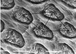
Enamel prism a structure characteristic of mammalian teeth, consisting of parallel bundles of enamel crystallites bounded by a cylindrical or hemicylindrical sheath of other crystallites which are likewise parallel, but oriented at a sharp angle to the bundle. The image shows fossilized enamel prisms from a multituberculate, Meniscoessus.
Enameloid a form of bone with even greater density and mineralization than dentine, usually found as a superficial layer over a dentine structure (e.g. scales of Paleozoic fish); mesodermal derivative laid down at outer surface of mesodermal papillae; may be up to 25% organic material.
Endochondral bone bone which is pre-formed as cartilage, as opposed to dermal bone.
Endocraniummore or less same as neurocranium, chondrocranium.
Endoderm The inner layer of tissue formed in the gastrula stage of development. At the end of blastula, the cells are arranged in the form of a hollow ball. Cell movement during gastrulation results in an invagination so that the embryo comes to resemble a double-walled cup. The inner layer of the cup is the endoderm. Endodermal cells usually end up forming the gut, pharynx, liver, lungs, and similar structures.
Endolymphatic duct in chondrichthyes and some other fishes, the inner ear has an external opening (or at least an opening outside the braincase) via an endolymphatic duct.
Endothelium the tissue lining circulatory or lymphatic vessels.
Ensenadan Age South American Land Mammal Age corresponding to the middle Pleistocene.
Entepicondylar foramen (or fossa) a hole (foramen) or groove (fossa) in the distal humerus, proximal to or on the entepicondyle which accommodates blood vessels and nerves associated with the humeral (ulnar) flexor muscles. See image under entepicondyle. See also Humerus.
Entepicondyle on the distal humerus, the condyle which faces posteriorly (for sprawling tetrapods) or medially (for erect tetrapods). See figure. This structure serves as the origin for the flexor muscles of the manus. See Humerus. With fine disregard for the hard facts of geometry, note that the entepicondyle faces outward in most illustrations, while the ectepicondyle protrudes between the two distal articulations of the humerus.
Enterocoely See Early Development Terms.
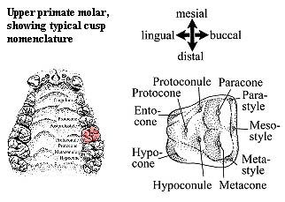
Entocone in mammalian upper molars. The nomenclature for small cusps in the mesiolingual (toward the tongue and anterior) region of upper molars is difficult. If the cusp is on the main body of the tooth, it is a protoconule. If it is a stylar cusp derived from the cingulum) it is a protostyle. If it takes the form of a ridge, it is an entocone. The figure shows an upper right molar with both an entocone and a protoconule. Note that the protoconule, if present, will lie along the crista (ridge) connecting the protocone and the paracone, if such a crista is present. In any case it will be somewhere along a theoretical line between the two and probably mesiobuccal to the protocone. An entocone or protostyle is likely to be further out on the margin and mesiolingual to the protocone.
Entoconid in mammalian dentition, a major cusp on the lingual side of the talonid (i.e. the linguodistal quadrant of the molar) in lower molars. See Molars.
Entocuneiform one of the distal tarsals. See ectocuneiform for image and explanation.
Entoplastron one of the dermal bones in the plastron of turtles.
Entopterygoid This is a frustrating term, as it seems to refer to 3 different and probably non-homologous bones: (a) a key hinge bone in the suspensorium of certain actinopterygian fishes cichlids?), (b) tooth-bearing plates flanking the parasphenoid in dipnomorph sarcopterygians, and (c) synonymous with the pterygoid in temnospondyls. However, such ambiguity is not without its uses. The next time you are confronted with some random fragment of vaguely palatal bone you may stroke your beard (or some more acceptably gender-neutral pilosity) and confidently pronounce, "Ah, yes. Of course. Undoubtedly a fragment of the entopterygoid." Then move on with your reputation for osteological omniscience untarnished. See also pterygoid.
checked ATW050621