| Palaeos: | Glossary | |
| The Vertebrates | Humerus |
| Page Back | Unit Back | Unit Home | Unit Dendrogram | Unit References | Glossary | Taxon Index |
| Page Next | Unit Next | Vertebrates Home | Vertebrate Dendrograms | Vertebrate References | Bones | Time |
"Let us go then, you and I
When the evening is spread out against the sky
Like a patient etherized upon a table"
T.S. Elliot, The Love Song of J. Alfred Prufrock
At the most simplistic level, the humerus is the bone which connects the shoulder to the elbow; and at the most refined level, it is the forelimb propodial. Somewhere in between, there is undoubtedly some level of jargon which is appropriate to the reader's own inclination and purposes. At a higher level of detail, the humerus has a remarkably complex topology -- one that has also changed dramatically over the course of tetrapod evolution. We cannot do it justice in the cramped format of a glossary entry. Accordingly, although this is still a far from adequate treatment, we will use this somewhat expanded space to provide a few schematics and landmarks. For a fuller treatment, see Romer (1956).
Initially, there is the matter of orientation. We will concentrate on the dorsal and ventral views. But what does "dorsal" mean with respect to a limb? By their nature, limbs do not have a fixed orientation with respect to the force of gravity. This problem is solved by adopting roadkill as a reference posture. To achieve this posture, one mentally splays the organism out on the center lane of Interstate 10, limbs straight out to the sides, and allows imaginary rush-hour traffic to do its worst. While this does little for the specimen quality of real animals, it quickly renders complex three-dimensional concepts as easily digested (perhaps a poor choice of words) two-dimensional shapes in dorsal view. Needless to say, quick application of a conceptual spatula generates the corresponding ventral view. Dorsal roadkill view is actually quite different from the orientation of tetrapods which do not sprawl. However, for purposes of comparability, we will use the same directional indications throughout. In tetrapods with an erect stance, the conventional directional indications are usually shown in parentheses.
Romer analyzes the humerus as the four triangular faces of a tetrahedron. This is excellent for basal tetrapods, and for those with samples and laboratory facilities. It is less easily applied to the flatter humeri of most Mesozoic forms, or by those without access to labs or to a teaching assistant (the T.A. is the pale, hungry-looking person with straggly, unkempt hair and a slightly frightening laugh who apparently lives under the lab bench). Nevertheless, it is well to remember that the humerus, primitively, is a four-sided object, and that we are collapsing it for pedagogical reasons.
 Basic
Features of an Early Humerus
Basic
Features of an Early HumerusThe figure at right illustrates some of the fundamental features of the left humerus of Bob in dorsal and ventral roadkill view. Fortunately, this is almost as bad as it gets. More derived tetrapods usually have much simpler humeri. However, Bob is our customary starting point. He is also Romer's starting point on the topic. So, 'though our choice be dubious, yet it is made on impeccable authority.
The most proximal point on most humeri is the proximal articulation which fits into the glenoid articulation formed by the scapula and coracoid. Primitively, this is an elongate curved surface which permitted the humerus to rotate anteriorly or posteriorly, allowed for a limited degree of motion up or down, but supported very little rotation around the long axis of the humerus. [1]
In some cases, we will find an even more proximal structure in the form of a lateral tubercle (sometimes referred to as the internal tuberosity), as in the aberrant bird (?) Mononykus or the turtle Platypeltis. This arises from the posterior margin of the humerus and sticks out at an angle, sometimes rising above the main proximal articular surface. However, it is infrequently encountered. The more typical homologous structures is the small, unmarked tubercle shown on the posterior margin of Bob's humerus, and the horizontal ridge on the posterodorsal surface, both just above the triceps attachment. These serve as attachments for muscles originating on or near the coracoid, or from the body wall. One relatively common example of the latter is the m. latissimus dorsi. In early tetrapods, such as Bob, the latissimus dorsi process is an easily recognizable feature of the dorsal humerus.
Another common structure in later tetrapods is the deltopectoral crest, which most typically occurs as a long ridge near the anterior edge of the proximal humerus, oriented generally with the long axis of the humerus. However, it may take other forms. Bob has no deltopectoral crest, as such, since he has a large, separate pectoral insertion on the ventral face of the upper humerus. However, one can already note the beginnings of a characteristic deltoid "tab" on the anterior margin in dorsal view.
The distal portion of the humeral shaft may be thought of as being divided into two epicondylar areas. The posterior moiety (or lateral moiety in graviportal and other non-sprawling forms) is the entepicondyle. In roadkill view, the large distal flange of the entepicondyle is usually a good landmark for early tetrapods and serves as an arrow pointing posteriorly. The entepicondyle is associated with the origin of the flexor muscles of the manus, the muscles which flex the wrist and close the fist. The entepicondyle is minimally scored by a groove to accommodate the passage of major blood vessels and nerves. Very frequently, this groove is roofed over and becomes a foramen, the entepicondylar foramen. Sadly these road signs are generally lost in more derived tetrapods.
 The anterior, or (primitively) the anterodorsal portion of the humerus is
likewise expanded as the ectepicondyle. In reality, i.e., in a
three-dimensional basal tetrapod, the ectepicondyle is actually oriented more
dorsally than anteriorly. The ectepicondyle is associated with the origin of the extensor musculature of the wrist and manus. Proximal
to this region, the ectepicondyle may merge with a more anteriorly-oriented supinator
process from which originate the muscles which rotate the wrist. The
ectepicondyle may also bear a groove or foramen, the ectepicondylar foramen.
The anterior, or (primitively) the anterodorsal portion of the humerus is
likewise expanded as the ectepicondyle. In reality, i.e., in a
three-dimensional basal tetrapod, the ectepicondyle is actually oriented more
dorsally than anteriorly. The ectepicondyle is associated with the origin of the extensor musculature of the wrist and manus. Proximal
to this region, the ectepicondyle may merge with a more anteriorly-oriented supinator
process from which originate the muscles which rotate the wrist. The
ectepicondyle may also bear a groove or foramen, the ectepicondylar foramen.
The distal surface of the humerus bears the articulations for the ulna and radius. The ulnar articulation is called a trochlea, although it frankly looks very little like the usual trochlea in the case of basal tetrapods. The ulnar trochlea allows the ulna to rotate principally only in one plane. The radial articulation is located more anteriorly and ventrally. It consistes of a rounded surface, the capitellum, which accommodates the broad range of motion necessary for rotation.
Rather than pretending that we took all this in at first intention, let us work through some examples. The reader is sternly warned that we have no answer book. Possible error lurks at every turn. Even in the professional literature, limb bones are frequently mislabeled or reversed. You have now been cautioned, and fore-warned is forearmed ...
 Example
#1
Example
#1In the first example, the entepicondyle is easily identified as a prominent tab of bone sticking out to the right of the distal end. Do not leap to the conclusion that this is therefore a humerus in dorsal view. It might be, for example, a right humerus. Look at the left side of the distal end. There are two rounded structures, one of which must be a capitellum. Therefore, our darkest fears are confirmed. This is a right humerus in ventral view. The entepicondylar foramen is unaccountably displaced anteriorly, compared to Bob's humerus. Another feature which suggests that the view is ventral is the overall concave shape of the proximal portion. This clue is not infallible, but frequently works, particularly for lepidosauromorphs. Yet another clue is the relative invisibility of the ectepicondyle, which is seen almost edge-on. If this is a ventral view, then the hook-shaped process on the anterior half of the proximal humerus is a pectoralis insertion, and not a latissimus dorsi process or triceps tubercle. Note that the posterior portion of the proximal end bears a small lateral tubercle. Click on the image to reveal the completely labeled figure.
 Example
#2
Example
#2The second example is considerably harder. The proximal articulation is round. Therefore we are dealing with a derived form in which the forelimbs are carried under the body. In these, later tetrapods, our conventional roadkill dorsal face is now posterior. The epicondyles and their foramina are no longer useful indicators. The epicondyles are reduced, flattened and roughly symmetrical. The epicondylar foramina are lost. Instead, we take our cue from the enormous deltopectoral crest The deltopectoral crest is roadkill anterior (lateral) and generally dorsal (posterior). Thus, this must be a left humerus in dorsal (posterior) view. The angle of the proximal articulation confirms this. The proximal articulation leans toward the right (medially) into the glenoid (articulation with the pectoral girdle), which confirms our fond expectations. Accordingly, the odd looking protuberance on the posterior (medial) side is probably a latissimus dorsi process.
An important point to note is that, with the transition to a more erect stance, everything rotates except the relative position of the radius and ulna (epipodials). In a sprawling stance, the epipodials are arranged along the line of the distal humerus with the radius anterior, and the ulna posterior. When the humerus rotates under the body, the radius is still anterior, not lateral. The ulna is still posterior, not medial. Now, however a line between them is transverse to the line of the distal humerus, rather than parallel.
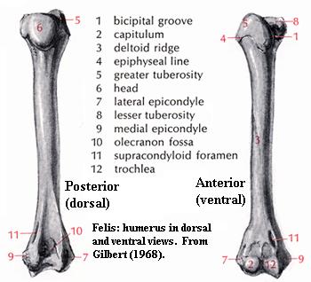 The
final example is not provided in self-test format because it cannot readily be
analyzed in terms of the landmarks provided above. Instead, we have simply
reproduced an illustration of a cat humerus from Gilbert
(1968), virtually without change. Note that a few of the basic
boundary stones remain. The capitellum (transformed by medical terminology
into the capitulum [2]) remains an easily-identified
feature of the distal end of the ventral humerus. On the dorsal side, the
distal humerus develops a long olecranon
fossa to accommodate the olecranon process, a proximal extension of the
ulna. However, the ulnar trochlea is changed into something resembling
much more closely a conventional pulley-like trochlea.
The deltopectoral crest is now found on the ventral face, and in a far more
distal position than we might expect. The proximal humerus is almost
unrecognizable with reference to Bob.
The
final example is not provided in self-test format because it cannot readily be
analyzed in terms of the landmarks provided above. Instead, we have simply
reproduced an illustration of a cat humerus from Gilbert
(1968), virtually without change. Note that a few of the basic
boundary stones remain. The capitellum (transformed by medical terminology
into the capitulum [2]) remains an easily-identified
feature of the distal end of the ventral humerus. On the dorsal side, the
distal humerus develops a long olecranon
fossa to accommodate the olecranon process, a proximal extension of the
ulna. However, the ulnar trochlea is changed into something resembling
much more closely a conventional pulley-like trochlea.
The deltopectoral crest is now found on the ventral face, and in a far more
distal position than we might expect. The proximal humerus is almost
unrecognizable with reference to Bob.
| Anterior (lateral) |
Posterior (medial) |
| Deltopectoral crest | Latissimus dorsi process |
| Ectepicondyle and/or supinator process |
Entepicondyle |
| Extensor & supinator muscles |
Flexor muscles |
| Capitellum | Trochlea |
| Radius (always anterior, never lateral) |
Ulna (always posterior, never medial) |
| Ulnare and pisiform (if present) |
Radiale and centralia (if any) |
| Digit V | Digit I |
To put the matter in context, and continuing in a highly over-simplified way, we may think of the arm in a slightly different way. Imagine the entire arm as having two tracks: anterior (lateral) and posterior (medial). (The directional indications in parentheses refer, as before, to the orientation in non-sprawling forms.) On the two tracks we have parallel landmarks, as shown in the table.
Because the epipodials do not rotate from their original basal positions, the radius can continue to articulate with the medial carpals, primarily the radiale; and the ulna continues to articulate with the lateral wrist bones, the ulnare and pisiform. However, this arrangement is devilishly hard to draw in an accurate and informative manner. Many sketches of the lower forelimb are, as a result, rather mysterious and vague.
It may be that this topic is simply too difficult for presentation without real bones and real troglodyte T.A.s. However, we hope it will serve as a useful introduction to an exceedingly difficult point of osteology. ATW020927.
[1] As an aside, the general trend in tetrapod locomotion has been to further develop those abilities in more or less that order, i.e. horizontal movement, vertical movement, and rotation about the long axis. For those with a biomechanical bent, you may try the following non-laboratory demonstration. Lie flat on the ground with arms straight out in roadkill position. (1) Move your arms forward and backward horizontally. These movements are, respectively, protraction and retraction. The angle of protraction or retraction is conventionally noted (presumably with a protractor or retractor) by the Greek letter theta, which looks like a zero in italics with a horizontal line through the middle. (2) If you are athletic enough, move your arms in, as in a push-up, and back out flat. These vertical movements are abduction and adduction. The abduction angle is conventionally noted by the Greek letter phi, which looks like zero with a vertical line through the middle. (3) Finally, rotate your arms about their long axes. This is, oddly enough, referred to as rotation, and is sometimes denoted by the Greek letter omega, a script 'w,' which may be imagined as a zero with the top half rotated down to the right.
[2] For once, the physicians have the right of it. Capitulum means "little head" in classical Latin. In later texts it also came to mean "chapter" or "heading" as in a book. Capitellum, by contrast, has a suspiciously Italianate sound to it, and may come from Renaissance anatomy texts. We owe an enormous debt to these anatomists, the first scientists in the modern sense. But, although their science was often excellent, their Latin was often not.
| Page Back | Unit Home | Page Top | Page Next |
checked ATW050828