|
|
Bones: The Braincase |
| The Vertebrates |
The Mastoid |
The Mastoid (Human Otic Capsule)
The mastoid is one of those repulsive little osteological features which mammals seem to have evolved solely for use as a badge of distinction, like a secret handshake or a fraternity code word. Indeed the possession of a particularly flagrant and obnoxious mastoid was once held to be the mark of man's superiority over the ape and the European's superiority over everyone else. By this reasoning, the world should rightfully be run by kangaroo rats, which have notably expanded "mastoids." But then, given the general direction of things, perhaps the world is being run by kangaroo rats.
Whether or not we are the pawns of xerophillic, megalomaniacal rodents bent on world domination, we must expose the truth, which is that the mastoid is not really anything new or different. It is simply another name for the periotic, which is identical to the petrosal, which is more or less the same thing as the otic capsule: i.e., the opisthotic plus the prootic. The "mastoid" is simply the homologous human variant of this structure. Accordingly, we will use this page as a place to summarize the status of the otic complex in human beings. In humans, the otic capsule has fused with the squamosal and various other odds and ends to form the temporal bone. The mastoid is then that portion of the temporal bone which encloses the middle ear and forms the outer wall of the inner ear in humans. It differs from the same structure in basal mammals and mammaliforms primarilly in its outer aspect. That is, it comes equipped with an outer ventral process, the mastoid process, to which a group of muscles attach. In humans, and a number of other mammalian taxa, the structure is also expanded and heavily pneumatized. This is said to have something to do with auditory acuity, but no one seems to know exactly what.
Anatomy & Attachments of the Otic Capsule in Humans
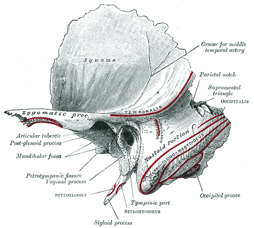 The figure on the right, lifted from the estimable Dr. Gray, shows the human left temporal bone in the sort of antero- latero- and- a- bit- dorsal view physicians use to talk down to patients without risking unseemly eye contact. The dorsolateral surface of the mastoid portion forms a shelf, sometimes referred to as the occipital ridge (since it lacks a ridge and is nowhere near the occiput). The shelf provides an attachment point for two vital muscles, the mm auricularis and occipitalis. These serve, respectively, to wiggle the ears and raise the eyebrows.
After this promising start, the mastoid shelf abruptly gives up, as if discouraged by such pointless activity, and drops down, flat and wrinkled, to a roughly rounded point ventrally. This is the mastoid process, named for its supposed resemblance to a breast. From this nomenclature we may charitably conclude that the medical anatomists responsible for the simile were perhaps unfamiliar with the secondary female characters of their own species. Attached to the mastoid process are the mm. sternocleidomastoideus, splenius capitis, and longissimus capitis. If one were, for example, to hang one's head sadly and slowly shake it back and forth as if ruefully contemplating some new and distressing demonstration of the perversity of fate, one would then be exercising all of these mastoid faculties.
The narrow space on the medial face of the mastoid process provides a shelter for the occipital artery, and also anchors the posterior body of the m. digastricus. This muscle inserts on the hyoid. Lifting the hyoid through the digastricus aids in swallowing and in opening the jaw. Note how easily the simple act of opening the mouth, while repeating the mastoid exercise described above, converts a sophisticated gesture of world-weary resignation into an expression of feckless and bovine stupidity.Surely there is some sort of lesson to be learned here.
Internally, the mastoid is hollowed out to form the epitympanic recess to accomodate the ossicles of the middle ear. At the blind end of this chamber, the bone forms the tegmen tympani. Actually, the tegmen is not a blind end, since it communicates with a complex of small air-filled chambers in the mastoid. As noted above, the function of these pneumatized pockets is unknown. ATW030519
checked ATW030506